Advertisement
Grab your lab coat. Let's get started
Welcome!
Welcome!
Create an account below to get 6 C&EN articles per month, receive newsletters and more - all free.
It seems this is your first time logging in online. Please enter the following information to continue.
As an ACS member you automatically get access to this site. All we need is few more details to create your reading experience.
Not you? Sign in with a different account.
Not you? Sign in with a different account.
ERROR 1
ERROR 1
ERROR 2
ERROR 2
ERROR 2
ERROR 2
ERROR 2
Password and Confirm password must match.
If you have an ACS member number, please enter it here so we can link this account to your membership. (optional)
ERROR 2
ACS values your privacy. By submitting your information, you are gaining access to C&EN and subscribing to our weekly newsletter. We use the information you provide to make your reading experience better, and we will never sell your data to third party members.
Biological Chemistry
Proteins From Birth To Death
From their biosynthesis to their folding, signaling, and degradation, proteins command the attention of many researchers
by Stu Borman
August 27, 2007
| A version of this story appeared in
Volume 85, Issue 35
PROTEINS ARE BORN on the ribosome, where they are biosynthesized. They may die in the proteasome, a big protein grinder in cells. And between those two events, "there is a life for proteins" in which they do many things, said biochemist Fabrizio Chiti of the University of Florence at a recent symposium. "They can carry out catalysis, they are involved in signal transduction, they help transport many other biomolecules, and they can act as genetic elements," among many other roles.
If one could understand everything that happens to proteins between the time they are born and the time they die, then one would indeed know everything there is to know about proteins. That is the ultimate goal of scientists who convened last month at the 21st Symposium of the Protein Society, in Boston. Chiti and biologist Judith Frydman of Stanford University were the symposium's program chairs.
In keeping with the meeting's theme, "Proteins: From Birth to Death," many presenters traced parts of the life story of proteins, from their biosynthesis on ribosomes to protein folding and from their functional existence in cells to their ultimate demise from degradation.
On the birth side of the spectrum were several talks on the way proteins come to life on ribosomes. Professor of molecular biology and genetics Rachel Green of Johns Hopkins University discussed her group's use of mutational and kinetic studies to unravel the way conformational rearrangements help the huge protein-RNA ribosomal complex do its job. Green and coworkers have recently been studying events that occur at the end of protein biosynthesis, when proteins called release factors bind to the ribosome and help it terminate the process in a precise and coordinated fashion.
The ribosome has two subunits, one small and one large. The small subunit is the place where the three-nucleotide anticodon of a transfer RNA binds in a highly specific manner to an amino acid-encoding sense codon on messenger RNA. It is also where the recognition site of a release factor binds just as specifically to a protein-terminating stop codon. In the large subunit, tRNA binding leads to peptide bond formation, which extends the growing protein one amino acid at a time. Also in the large subunit, binding of a release factor results in a hydrolytic reaction that terminates protein translation.
Release factors not only recognize a stop codon with high fidelity in the small subunit, but they also direct the ribosome's peptidyltransferase catalytic site to use a water molecule and thereby end the protein chain, instead of using an amino acid and thereby extend the chain. Green and coworkers have shown that high-fidelity stop-codon recognition in the small subunit and activation of the peptidyltransferase center in the large subunit both occur by induced-fit mechanisms, which are conformational changes that serve as prerequisites to other actions.
Binding of release factor to a correct stop codon induces ribosomal shape modifications that send a signal allowing hydrolysis to occur in the peptidyltransferase center. tRNAs also use induced-fit mechanisms for codon recognition and peptidyltransferase activation during protein elongation.
Green and coworkers have found new details about how these processes occur. "It appears that the ribosome functions by responding to a series of switches in the presence of the correct substrate, either tRNA or release factor," Green said. "A correct codon-anticodon or stop-codon/release-factor interaction triggers a switch in the small subunit, and then a subsequent switch is induced in the large subunit to trigger catalysis."

AFTER A PROTEIN is born, it has to fold properly to be able to go to work in the cell. Proteins fold in the endoplasmic reticulum (ER) before being moved to other locations where they are needed. If the ER has inadequate capacity to handle all the proteins that are being produced and develops a buildup of unfolded proteins, it uses a feedback circuit, called the unfolded protein response, to alert the nucleus about the problem. At the symposium, University of California, San Francisco, biochemistry and biophysics professor and Howard Hughes Medical Institute investigator Peter Walter told attendees about progress in understanding the mechanism of the unfolded protein response.
In yeast, components of the unfolded protein response include IRE1, a bifunctional transmembrane enzyme. Binding of an unfolded protein in a pocket on the end of IRE1 inside the ER may tether two or more IRE1s together. The tethering causes IRE1's kinase domains on the other side of the membrane to become phosphorylated and to bind nucleoside phosphates such as adenosine triphosphate (ATP).
The action in the kinase domains, in turn, causes a conformational change that enables IRE1 to exhibit another function: It becomes a highly specific endoribonuclease whose activity, splicing the mRNA of the transcription factor HAC1, results in signals to the nucleus to increase the ER capacity of the cell.
In mammalian cells, IRE1 performs a similar splicing reaction on the mRNA of a mammalian transcription factor called XBP1. The actions of IRE1 and other ER transmembrane proteins like PERK and ATF6 result in signals being sent to the nucleus to adjust the ER's protein-folding capacity to accommodate cellular needs.
Also in mammalian cells, the relative timing of signaling by IRE1, PERK, and ATF6 may lead to life-and-death decisions. If the capacity of the ER can't be adjusted, and too many unfolded proteins continue to accumulate, the unfolded protein response turns on a cell-suicide (apoptosis) pathway.
This life-and-death decision-making ability makes the unfolded protein response potentially relevant to various diseases. In retinitis pigmentosa, inherited mutations in rhodopsin molecules cause improperly folded rhodopsin to accumulate in the ER, leading to apoptosis of rod cells and consequent blindness. Other inherited protein-folding diseases, as well as diabetes, cancer, and viral infection, may also be affected by the unfolded protein response. "We want to better understand the mechanism of the unfolded protein response to find rational ways of affecting these processes and thereby improve human health," Walter said.
As if accumulations of unfolded proteins aren't bad enough, problems can also arise when proteins misfold. For example, misfolded proteins called prions can cause disease, and their quick detection has been an important research goal.
Prions are infectious proteins that cause transmissible spongiform encephalopathies (TSEs), protein-misfolding and -aggregation diseases such as mad cow disease in cattle and Creutzfeldt-Jakob disease in humans. New prions form when normal prion protein, an endogenous protein found on neuron membranes, is exposed to existing prions and thereby induced to misfold—the way a rough kid at school might convince a classmate to join a gang.
It's essential to be able to detect a TSE as soon as possible after an animal or person gets the disease. Early detection can prevent infected cows from being processed into meat and other products that might spread the disease to animals and people. And early detection in people opens the possibility of more effective therapeutic intervention, although no therapies for Creutzfeldt-Jakob disease have yet been validated in clinical trials.
A few years ago, a group led by neurology professor Claudio A. Soto, now at the University of Texas Medical Branch, in Galveston, developed a technique called protein misfolding cyclic amplification (PMCA), which greatly increases the concentration of prions in a sample so they can be detected more easily. In PMCA, a prion-containing sample from brain or other tissue is used as a seed to induce misfolding of normal prion protein. The misfolded protein forms aggregates, which are sonicated to maximize the surface area of misfolded seed material. Conversion and sonication are repeated until the prions in the original sample have been amplified millions of times.
PMCA is extremely sensitive and is faster than conventional bioassays. Like current bioassays, however, it requires brain samples, which are difficult to obtain; it also takes three weeks or more for PMCA to be carried out with optimal sensitivity.
A better way to detect the proteins has come from a group led by senior investigator Byron Caughey of the National Institute of Allergy & Infectious Diseases' Rocky Mountain Laboratories, Hamilton, Mont. Caughey and coworkers devised a variation on PMCA called recombinant prion protein PMCA (rPrP-PMCA). Compared with conventional PMCA, it has 10- to 50-fold lower sensitivity—a level still adequate for disease detection—but it doesn't require brain samples and is much faster (Nat. Methods 2007, 4, 645).
rPrP-PMCA uses recombinantly derived normal prion protein as the amplification source material instead of the small amounts of normal prion protein in brain samples used for conventional PMCA. This enables rPrP-PMCA to detect prions in cerebrospinal fluid samples, which are much easier to obtain than brain samples, and allows detection in only three days instead of 21 or more. Caughey noted that in addition to its assay and diagnostic potential, rPrP-PMCA could be used to advance fundamental research on the structure of infectious prions and the mechanism of their formation.
In addition to detecting proteins, researchers would like to visualize them better, and a Texas-based team has just demonstrated improved capabilities of an electron microscopy-based method for protein structural analysis.

Single-particle cryoelectron microscopy (cryo-EM) is a technique for imaging molecular complexes immobilized in a thin layer of ice. Cryo-EM can determine samples at low concentrations. It doesn't require that complexes be crystallized, which can be difficult or impossible to achieve. And it can analyze different conformations instead of being locked into a single crystallized form. Therefore, cryo-EM is being developed as an alternative to crystallography for determining structures of protein complexes.
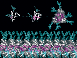
From tens of thousands of cryo-EM images of single particles of the large, 800-kilodalton GroEL chaperonin complex, structural biologist Wah Chiu and coworkers at Baylor College of Medicine, in Houston, put together a high-resolution structure through image reconstruction. They have been able to trace the polypeptide backbone of the biomolecular machine without any reference whatsoever to its X-ray crystal structure. This is the first time an entire polypeptide backbone has been traced with single-particle cryo-EM, Chiu said, demonstrating the improving prospects of cryo-EM for biomolecular structural analysis.
Natural proteins are so good at the tasks they perform that sometimes the best compliment researchers can give them is to emulate their functional powers with designed versions. Yale University postdoc Tijana Z. Grove, molecular biophysics and biochemistry professor Lynne J. Regan, grad student Jason Forster, and mechanical and chemical engineering professor Eric Dufresne have done just that by engineering protein-based biomaterials for potential biomedical applications.
They have been focusing on the design and engineering of tetratricopeptide repeat proteins (TPRs) that can recognize specific biological motifs. TPRs are naturally occurring 34-amino-acid peptides that adopt helix-loop-helix structures. Sets of three to 22 TPRs can be combined in a modular fashion to create stacked structures, which can be incorporated into biomaterials. These TPR-based biomaterials have potential applications in wound healing, tissue regeneration, and sensing; the Yale researchers have focused on drug delivery.
The team used TPRs to form porous hydrogels. The materials can be dissolved by using low-pH conditions or specific small molecules to inhibit binding interactions that are essential to their integrity. If the hydrogels are engineered to contain drug molecules, the drug will be released by hydrogel dissolution.
TPRs in the hydrogel-based drug delivery systems can be designed to recognize cell receptors, such as the HER2 growth factor receptors on cancer cells. Binding to such receptors enables the hydrogels to be internalized only by cancer cells. The low pH inside the cells then causes the hydrogels to dissolve and release their anticancer-drug cargo exactly where it's most needed, Grove said.

IN CASE the right protein for a specific job isn't available, computational methods can be used to redesign proteins to have useful new properties. University of California, San Diego, professor of pharmacy Tracy M. Handel and coworkers recently used a program they developed to redesign interactions between a key bacterial enzyme and an inhibitor protein that binds to it.
Advertisement
The program, called EGAD Library (J. Comput. Chem., DOI: 10.1002/jcc.20727) uses mostly theoretical principles and a minimal amount of experimental data to fairly accurately predict the stabilities of protein mutants and protein complexes. Handel and coworkers used the program to redesign complexes of β-lactamases with β-lactamase inhibitor protein (BLIP).
β-Lactamases catalyze the reactions that render β-lactam antibiotics like cephalosporin and penicillins inactive and thereby contribute to the growing problem of bacterial resistance to antibiotics. Because BLIP binds and inhibits β-lactamases, it's a model for drugs that could lower bacterial resistance.
BLIP has a range of affinities for different binding partners. For example, it binds to β-lactamase TEM-1 more than 1,000 times tighter than it binds to β-lactamase SHV-1, even though the two β-lactamases are structurally nearly identical. By analyzing the two interactions, Handel and coworkers were able to make BLIP bind to a modified SHV-1 as strongly as it binds TEM-1 by changing a single amino acid in the SHV-1 binding site.
They then used EGAD Library to redesign BLIP itself to overcome its low affinity for native SHV-1. The modified BLIPs bind native SHV-1 with much higher affinity than wild-type BLIP does.

ALSO ALONG the lines of protein modeling, another research team is examining the properties evolutionarily unimproved proteins might have had by creating a primitive proteome. Researchers have long proposed that primitive enzymes might have been capable of reacting with a wide range of substrates, because that would have optimized the catalytic versatility of ancestral cells possessing a limited catalytic repertoire. But it's difficult to test the idea, because all enzymes today have already been subjected to evolution.
So grad student Shona C. Patel, postdoc Luke Bradley, and chemistry professor Michael H. Hecht at Princeton University used protein design to make a primitive proteome of simple protein structures. They created about 1 million diverse sequences designed to fold into four-helix bundles, with nonpolar residues inside and polar residues outside, as in natural proteins. Then they expressed the proteins in bacteria. They found that several proteins in a randomly selected group shared four functional properties: heme binding and peroxidase, esterase, and lipase activities. The results show that "naive" proteins can indeed adopt the type of wide-ranging activity that has been proposed for primitive enzymes.
Other protein research projects discussed at the symposium included studies on disordered proteins and the effect of proteins on biological evolution.
Intrinsically disordered proteins contain unstructured regions under physiological conditions (C&EN, April 2, page 58). For a very long time, they were "completely overlooked by the scientific community," said Vladimir N. Uversky, senior research professor of biochemistry and molecular biology at Indiana University School of Medicine. "This was because of the predominating viewpoint that a protein is a highly organized and rigid molecule with a unique structure specially designed for a unique function."
About a decade ago, however, researchers discovered that partly or even fully unfolded proteins could still be functional. Uversky and coworkers recently surveyed the functionality of intrinsically disordered proteins and their strong association with human diseases (J. Proteome Res. 2007, 6, 1882, 1899, and 1917). "Their most prominent functions are signaling, regulation, and recognition," he explained. "They are also very abundant in diseases like cancer, Alzheimer's disease, Parkinson's disease, prion diseases, cardiovascular diseases, and diabetes" and thus represent a new class of drug targets.
Intrinsically unfolded proteins present drug discovery with a new set of challenges, because drugs aren't likely to bind to or interact with their unstructured regions in the same way they do with folded protein domains. "Traditional approaches for drug discovery and design will not work with these proteins," Uversky said. "A new paradigm is being developed."
At Harvard University, meanwhile, a group is using ab initio protein modeling in a unique way: to provide a first-principles model of biological evolution. Professor of chemistry and chemical biology Eugene I. Shakhnovich and coworkers are preparing a new model of early evolution that directly relates the fitness of populations of evolving model organisms to the properties of their proteins (PLoS Comput. Biol., DOI:10.1371/journal.pcbi.0030139).
Shakhnovich and his coworkers have proposed that death rates of organisms are determined by the stability of their least stable proteins. And they find that survival and growth of a population is greatly enhanced after a small number of advantageous protein structures suddenly appear on the scene. The protein universe expands as such innovative proteins duplicate and diverge.
They also propose a speed limit on molecular evolution: no more than six mutations per genome for each replication of an organism. "At higher mutation rates, populations experience mutational meltdown; they go extinct due to failure of their proteins to fold," Shakhnovich said.
"Science is important for a number of reasons-for advancing our knowledge, for technical progress, curing our diseases, and many other things," said symposium organizer Chiti in his address to conferees. And learning more about proteins and how they live and work is an important part of all that. It's an effort that protein scientists engage in every day, as they continue to strive to understand the things proteins do at birth, at death, and at every stage in between.
You might also like...


The power is now in your (nitrile gloved) hands
Sign up for a free account to get more articles. Or choose the ACS option that’s right for you.
Already have an ACS ID? Log in
Join the conversation
Contact the reporter
Submit a Letter to the Editor for publication
Engage with us on Twitter