Advertisement
Grab your lab coat. Let's get started
Welcome!
Welcome!
Create an account below to get 6 C&EN articles per month, receive newsletters and more - all free.
It seems this is your first time logging in online. Please enter the following information to continue.
As an ACS member you automatically get access to this site. All we need is few more details to create your reading experience.
Not you? Sign in with a different account.
Not you? Sign in with a different account.
ERROR 1
ERROR 1
ERROR 2
ERROR 2
ERROR 2
ERROR 2
ERROR 2
Password and Confirm password must match.
If you have an ACS member number, please enter it here so we can link this account to your membership. (optional)
ERROR 2
ACS values your privacy. By submitting your information, you are gaining access to C&EN and subscribing to our weekly newsletter. We use the information you provide to make your reading experience better, and we will never sell your data to third party members.
Biological Chemistry
Fertilization Illuminated
Crystallography offers peek at a mammalian sperm receptor
by Carmen Drahl
December 8, 2008
| A version of this story appeared in
Volume 86, Issue 49

BY TAMING a tough-to-handle protein, researchers have provided a rare atomic-level glimpse at mammalian fertilization. Their X-ray crystal structure, reported in Nature (2008, 456, 653), may lead to new contraceptives, as well as further the understanding of reproductive and other ailments.
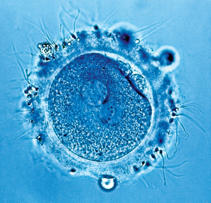
Fertilization begins when a sperm cell makes contact with sperm receptors on the outer coating of an egg cell. In human and other mammalian eggs, that coat, called the zona pellucida (ZP), consists of long fibers made of glycoprotein polymers. Some of the glycoproteins, called ZP domain proteins, also act as sperm receptors. The proteins' dense tangle of sugars and natural tendency to aggregate into filaments have confounded previous structural efforts. Now, structural biologist Luca Jovine and coworkers at Sweden's Karolinska Institute have sidestepped both of those problems and obtained the X-ray structure of an important fragment of a mouse sperm receptor.
Sugar-rich proteins are notoriously tough to crystallize. Rather than stripping the sugars away, Jovine's lab opted to study the 11-kilodalton amino-terminal fragment of the mouse sperm receptor ZP3. In mice, this part of ZP3 contains no sugars. The team preferred not to "wonder whether removing the sugars would still give a realistic structure," Jovine explains. To avoid aggregation, the team fused the ZP3 fragment to a bacterial protein. By adjusting the linker between the two proteins, they minimized aggregate formation and obtained crystals.

The protein portrait reveals an interaction between a pair of tyrosine residues, at least one of which is essential for polymerization and, therefore, egg coat formation. Furthermore, the team's molecular modeling studies suggest a mechanism by which the egg coat prevents more than one sperm from entering the egg. The Swedish team is now trying to obtain a complete structure of mouse ZP3.
The work provides "a great leap forward in our understanding of sperm reception by mammalian eggs," says Victor D. Vacquier of Scripps Institute of Oceanography, who has determined structures of invertebrate reproductive proteins. Learning about other mammals' sperm receptor structures could illuminate why sperm-to-egg binding is species specific, he adds.
"Defects in sperm interaction with ZP are linked to male infertility," says Harvey M. Florman, an expert in the mechanisms of fertilization at the University of Massachusetts Medical School, Worcester. The close-up view this structure provides could help explain what goes wrong in those cases, as well as lead to the design of small-molecule contraceptives that selectively block the sperm receptor, he notes.
Jovine's work has an impact beyond the fertilization field, adds Paul M. Wassarman, a ZP domain protein specialist at Mount Sinai School of Medicine, in New York City, who wrote a Nature commentary accompanying the report. ZP domains are found in filament-forming proteins in the human kidney, the inner ear, and elsewhere, and the structure could help researchers understand diseases in these organs, Wassarman says. For example, a mutation in one of the key tyrosines is associated with deafness, Jovine points out.
Advertisement
You might also like...


The power is now in your (nitrile gloved) hands
Sign up for a free account to get more articles. Or choose the ACS option that’s right for you.
Already have an ACS ID? Log in
Join the conversation
Contact the reporter
Submit a Letter to the Editor for publication
Engage with us on Twitter