Advertisement
Grab your lab coat. Let's get started
Welcome!
Welcome!
Create an account below to get 6 C&EN articles per month, receive newsletters and more - all free.
It seems this is your first time logging in online. Please enter the following information to continue.
As an ACS member you automatically get access to this site. All we need is few more details to create your reading experience.
Not you? Sign in with a different account.
Not you? Sign in with a different account.
ERROR 1
ERROR 1
ERROR 2
ERROR 2
ERROR 2
ERROR 2
ERROR 2
Password and Confirm password must match.
If you have an ACS member number, please enter it here so we can link this account to your membership. (optional)
ERROR 2
ACS values your privacy. By submitting your information, you are gaining access to C&EN and subscribing to our weekly newsletter. We use the information you provide to make your reading experience better, and we will never sell your data to third party members.
Analytical Chemistry
Nanostructure Dynamics
Imaging: Time-resolved electron tomography provides 3-D views on ultrafast timescale
by Mitch Jacoby
June 28, 2010
| A version of this story appeared in
Volume 88, Issue 26

By integrating time resolution into a method that generates three-dimensional electron microscopy images, researchers in California have developed a technique that provides a 3-D view of nanometer-scale specimens moving on the femtosecond timescale (Science 2010, 328, 1668). The procedure, dubbed 4-D electron tomography, offers a novel way to probe the dynamics of microscopic objects undergoing subtle transient motions and structural changes.
Conventional transmission electron micrographs are 2-D projections of 3-D specimens. By recording many 2-D projections at various viewing angles, which can be carried out by tilting the specimen incrementally and recording an image at each tilt angle, a 3-D composite image can be constructed with the help of computer algorithms. This well-established electron tomography procedure can provide researchers with detailed 3-D views of complex specimens from multiple perspectives. Although this type of analysis readily leads to geometric and structural insights that cannot be derived from flat projections alone, the information obtained in this way paints a time-averaged picture of a static object in an equilibrium state.
Now, such high-resolution electron tomography has taken on a new dimension: time. California Institute of Technology senior postdoc Oh-Hoon Kwon and chemistry Nobel Laureate Ahmed H. Zewail have developed a way to produce tomograms with nanoscale spatial resolution and femtosecond temporal resolution.
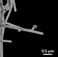
In a demonstration study, the team generated tomographic images and videos that capture a braceletlike carbon nanotube structure wiggling and “breathing” in response to sudden heating pulses. The method, which exploits laser-driven ultrafast electron microscopy technology pioneered by Zewail’s group, provides up-close views of a complex dynamic specimen and promises to have wide-ranging applications in biological and materials science, the team says.
“This is a stunning development in ultrafast time-resolved structural imaging,” says Charles M. Lieber, a chemistry professor at Harvard University. He adds that the Zewail group’s seminal demonstration of time-resolved electron tomography provides researchers with movies of the full 3-D atomic-level structure and dynamics of a nanostructure for the first time. This procedure represents “a remarkable new capability for nanoscience and will also undoubtedly impact substantially the life sciences,” he says.
Rice University chemistry professor James L. Kinsey is equally enthusiastic about the new imaging technique. “This work is an impressive extension of technology for studying structural change in nanoscale objects,” he says. “It would be hard to predict the impact of this advance on molecular science, but it is sure to be profound.”
Time-resolved electron tomography captures this carbon nanotube structure undergoing ultrafast morphological changes from various viewing angles.


Join the conversation
Contact the reporter
Submit a Letter to the Editor for publication
Engage with us on Twitter