Advertisement
Grab your lab coat. Let's get started
Welcome!
Welcome!
Create an account below to get 6 C&EN articles per month, receive newsletters and more - all free.
It seems this is your first time logging in online. Please enter the following information to continue.
As an ACS member you automatically get access to this site. All we need is few more details to create your reading experience.
Not you? Sign in with a different account.
Not you? Sign in with a different account.
ERROR 1
ERROR 1
ERROR 2
ERROR 2
ERROR 2
ERROR 2
ERROR 2
Password and Confirm password must match.
If you have an ACS member number, please enter it here so we can link this account to your membership. (optional)
ERROR 2
ACS values your privacy. By submitting your information, you are gaining access to C&EN and subscribing to our weekly newsletter. We use the information you provide to make your reading experience better, and we will never sell your data to third party members.
Analytical Chemistry
Multiplexed Raman microscopy
Tunable filter expands Raman imaging technique to multiple wavelengths simultaneously
by Celia Henry Arnaud
March 5, 2012
| A version of this story appeared in
Volume 90, Issue 10
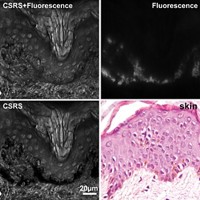
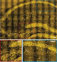
A new version of stimulated Raman scattering (SRS) microscopy speeds up image acquisition by allowing multiple bands to be measured simultaneously (J. Am. Chem. Soc., DOI: 10.1021/ja210081h). Dan Fu, Xiaoliang Sunney Xie, and coworkers at Harvard University and the University of Notre Dame achieve the multiplexing by adding an acousto-optic tunable filter (AOTF) to their SRS setup. In the AOTF, acoustic waves sent through a crystal diffract the broadband femtosecond pump laser beam into a number of bands, each corresponding to a Raman shift modulated at a different frequency. The filtered pump beam is combined with a picosecond laser beam and sent to a laser scanning microscope. The Raman shift is determined from the energy difference between the filtered pump beam and Stokes beam, which is encoded in the modulation frequency. The researchers use a Fourier transform to extract multiple Raman bands by demodulating the signal at the different frequencies. Using the method, the researchers measured the biochemical composition of microalgae. They used three channels to distinguish among pigments, lipids, and proteins in the cells. They also obtained images of blood, lipids, and proteins in freshly excised mouse ear tissue.


Join the conversation
Contact the reporter
Submit a Letter to the Editor for publication
Engage with us on Twitter