Advertisement
Grab your lab coat. Let's get started
Welcome!
Welcome!
Create an account below to get 6 C&EN articles per month, receive newsletters and more - all free.
It seems this is your first time logging in online. Please enter the following information to continue.
As an ACS member you automatically get access to this site. All we need is few more details to create your reading experience.
Not you? Sign in with a different account.
Not you? Sign in with a different account.
ERROR 1
ERROR 1
ERROR 2
ERROR 2
ERROR 2
ERROR 2
ERROR 2
Password and Confirm password must match.
If you have an ACS member number, please enter it here so we can link this account to your membership. (optional)
ERROR 2
ACS values your privacy. By submitting your information, you are gaining access to C&EN and subscribing to our weekly newsletter. We use the information you provide to make your reading experience better, and we will never sell your data to third party members.
Analytical Chemistry
CARS Microscopy, A New Window Into Geochemistry
An established biomedical imaging technique makes its debut in rock analysis
by Mitch Jacoby
November 19, 2012
| A version of this story appeared in
Volume 90, Issue 47

Geologists don’t usually attend biomedical imaging workshops. And if they do show up, they don’t typically come with rock samples in hand. But that’s exactly what Robert C. Burruss once did.
Burruss knew he would be the odd man out two years ago at a gathering of scientists who had come to learn about a Raman microspectroscopy method known to be a powerful technique for imaging fine details of cellular organelles. “Everybody thought I was crazy showing up with rock samples,” he says. But Burruss, a research geologist with the U.S. Geological Survey (USGS), wasn’t deterred.
He wondered whether that Raman method, coherent anti-Stokes Raman scattering (CARS) microscopy, could be tailored to uncover subtleties of geochemical processes associated with oil and gas trapped in rocks. Such information can be helpful in locating and characterizing buried petroleum reserves. So after discussing the seemingly odd idea with Albert Stolow, one of the workshop hosts and organizers and a laser scientist at the Canadian National Research Council (NRC) in Ottawa, Ontario, the USGS scientist headed to the workshop in Stolow’s lab with a handful of rock samples.
Before the brief workshop ended, the pair saw the first hints suggesting that, indeed, CARS microscopy might be able to probe hidden geochemical features, and they agreed to team up to further explore that possibility. Now the team has published the first study describing the proof-of-concept application of CARS microscopy to analyze trapped gases in geologic samples (Geology, DOI: 10.1130/g33321.1). And earlier this month, Stolow presented results of the study in Charlotte, N.C., at the annual meeting of the Geological Society of America.
Much of the original CARS microscopy development was done by a team led by Harvard University’s X. Sunney Xie. The method was recognized as a potentially powerful imaging tool, but one that required advanced laser skills. Stolow and colleagues at NRC developed a simplified variation of the method, and that advance led to the first commercial CARS microscope, which was debuted in 2009 by Olympus.
The microscopy technique is based on Raman spectroscopy, but it generates analytical signals four to five orders of magnitude more intense than conventional Raman spectroscopy, Stolow explains. Those properties enable researchers to quickly image tiny intracellular organelles without using microscopy stains or dyes by highlighting chemically distinct features of the organelles, such as fingerprint-like C–H vibrations in lipids.
That analytical prowess in biological imaging led Burruss to wonder whether CARS microscopy might prove useful in studying organic matter in “fluid inclusions.” Those geological features are micrometer-sized pockets in mineral crystals that contain long-ago-trapped fluids such as hydrocarbon oil and gas.
By understanding the geological processes that formed inclusions in a given rock sample and by knowing the chemical composition of the inclusions’ contents, scientists can determine much about the geological environment from which the sample was collected. For example, methane-rich inclusions in a quartz sample retrieved from a surface outcrop may indicate that the location contains a much more deeply buried shale-based natural gas reservoir.

Burruss had been using conventional Raman spectroscopy, microthermometry, and other methods to analyze inclusions for more than 25 years. But those standard geology methods have key shortcomings, he says. For example, thermometry provides phase behavior results that are not molecule-specific for complex fluids such as crude oil. And Raman data, although molecule-specific, are often inaccessible because they end up buried by the overwhelming fluorescence background caused by the presence of petroleum liquids. For those reasons and others, geologists are always on the lookout for improved ways to analyze organic matter in rocks.
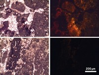
It wasn’t obvious at the outset that the CARS system in Stolow’s lab would generate much signal from rock samples. But when the researchers conducted a test run, “the sample lit up like a lightbulb,” Burruss says. “It was a really exciting moment.”
Energized by the results of the trial run, Stolow, Burruss, and two physicists, Aaron D. Slepkov of Trent University and Adrian F. Pegoraro of Queen’s University, both in Ontario, teamed up to develop methodology for applying CARS microscopy to analysis of fluid inclusions. The team has now demonstrated that the new method has significant advantages and unprecedented capabilities compared with standard geochemical analysis methods.
For example, the CARS signal rapidly reveals the three-dimensional distribution of methane-rich inclusions and pinpoints the presence of higher hydrocarbons without fluorescence interference, even when the methane is dissolved in brightly fluorescing crude oil droplets. Also, because of the way the measurements are made, the method simultaneously provides two-photon fluorescence data and second-harmonic generation (SHG) data, in addition to the CARS signal, from the sample volume under interrogation. The fluorescence image can be used to identify aromatic hydrocarbons, and SHG analysis provides crystallographic information.
That combination of data emanating from a microscopic point enables researchers for the first time to correlate the chemical composition of inclusions with detailed knowledge of their 3-D crystallographic microstructures. The method may also reveal other details such as the methane concentration and pressure inside gas- and liquid-filled inclusions.
“This work is a major contribution to geology,” says Robert J. Bodnar, a professor of geosciences at Virginia Tech. “To a geologist, a rock is like a book,” Bodnar says. In much the same way that readers try to develop a broad vocabulary to understand every word in a book, geologists work to develop analytical methods that help them “read” a rock’s history, he explains. “This new method allows us to analyze a range of materials that we could not analyze in the past and will help us learn about their history.”
Geologists aren’t the only ones excited about the study. Eric O. Potma, a chemistry professor and laser imaging specialist at the University of California, Irvine, finds it “amazing” that this method provides a way to extract chemical information from a tiny, hermetically sealed microscopic volume without physically touching, let alone destroying, the sample.
Potma adds, “The group has convincingly shown that this noninvasive form of chemical analysis, which has already found applications in biomedical imaging, offers an invaluable diagnostic for geological materials.”


Join the conversation
Contact the reporter
Submit a Letter to the Editor for publication
Engage with us on Twitter