Advertisement
Grab your lab coat. Let's get started
Welcome!
Welcome!
Create an account below to get 6 C&EN articles per month, receive newsletters and more - all free.
It seems this is your first time logging in online. Please enter the following information to continue.
As an ACS member you automatically get access to this site. All we need is few more details to create your reading experience.
Not you? Sign in with a different account.
Not you? Sign in with a different account.
ERROR 1
ERROR 1
ERROR 2
ERROR 2
ERROR 2
ERROR 2
ERROR 2
Password and Confirm password must match.
If you have an ACS member number, please enter it here so we can link this account to your membership. (optional)
ERROR 2
ACS values your privacy. By submitting your information, you are gaining access to C&EN and subscribing to our weekly newsletter. We use the information you provide to make your reading experience better, and we will never sell your data to third party members.
Materials
Understanding Joint Wear And Tear
Scientists decode surface phenomena that are key to extending implant life
by Mitch Jacoby
August 4, 2014
| A version of this story appeared in
Volume 92, Issue 31

John B. Medley’s interest in the wear and tear of hip replacements isn’t strictly academic. True, the University of Waterloo mechanical engineering professor heads a research group that studies friction, wear, and lubrication of joint implants in the laboratory. But for decades, Medley has even been carrying around an implant outside the lab—in his hip.
A motorcycle accident in 1975 badly dislocated Medley’s hip joint. “It was quite a traumatic accident,” he says. “I was lucky to have lived through it.”
Doctors managed to put the young scientist’s hip back together, buying him 10 or so reasonably trouble-free years. But eventually, osteoarthritis and related joint deterioration necessitated a hip replacement. Unfortunately, roughly 10 years later, surgeons needed to revise Medley’s implant, replacing worn prosthesis components with new ones.
This scenario is common. According to the American Academy of Orthopaedic Surgeons, the combined number of hip and knee replacements performed each year by doctors in the U.S. now sits at about 1 million. About 12% of those patients will likely undergo follow-up surgery in 10 years as a result of some type of implant failure or problem and associated complications.
The exact reasons for those failures are often unclear. Body joints are multicomponent interfaces with surfaces that undergo relative sliding, rotating, and flexing motions. The mechanics of those motions are influenced by a combination of processes that include the effects of wear, lubrication, chemical corrosion, and cellular response. The problem is complex. But with the number of patients undergoing joint replacement surgeries—just in the U.S.—predicted to exceed 4 million per year by 2030, materials scientists, medical doctors, engineers, and other researchers are driven to uncover the fundamental mechanisms that underpin wear, tear, and corrosion of joint implants.
For more than a century, doctors have been replacing body joints to relieve pain and restore function to patients whose joints have been destroyed by trauma or disease. Construction materials for artificial joints have evolved over the years and have included metals and alloys, synthetic polymers, and ceramics.
Among the key factors guiding choice of materials for joint replacements are the durability and friction of the implant components. In hips, for example, the top of the thigh bone (femur) is a ball-shaped structure that fits snugly in the pelvic bone socket (acetabulum). In healthy people, the relative motions of the bones in that joint generate little friction because the bones are protected and cushioned by a slippery cartilage layer. In artificial joints, frictional forces can be hundreds of times higher. Those forces lead to wear on the sliding surfaces and thus limit device longevity and can elicit adverse reactions in the bone and tissues surrounding the implant.
Hip replacement procedures caught on strongly with the introduction of a metal-on-polymer prosthesis design in the 1960s that featured a ball-shaped metal femoral head that fit into a polymeric acetabular cup. At first, the design seemed highly successful. But eventually, rubbing motions of the hard metal surface against the cup released micrometer-sized polymer particles into the joint area. In some patients, that process triggered osteolysis, a painful condition involving loss of bone mass that can cause implants to become loose. In some cases, it led to bone fractures in the implant area.

To minimize the release of harmful wear particles, biomedical device manufacturers began making metal-on-metal prostheses, with each of the rubbing surfaces often made from a cobalt-chromium-molybdenum alloy. According to Philippa Cann, a principal research fellow at Imperial College London, the metal-on-metal design exhibited excellent wear characteristics in lab tests and was expected to provide long implant service life. But soon it was found that these devices also deteriorated from abrasive rubbing, releasing biologically active nanometer-sized metal particles in the joint area.
“In some cases, however, these artificial joints functioned well for 30 years,” Cann says. “The right hip in the right person can work beautifully. But we still don’t know why.”
With unexpectedly high failure rates for metal-on-metal devices, manufacturers looked for better materials options. Many hip implants made currently include low-wear ceramic components. And metal-on-polymer implants have recently made a big comeback with a notable enhancements in material properties: Unlike the polyethylene of a few decades ago, the polyethylene found in today’s implants is a highly cross-linked ultra-high-molecular-weight material.
Several studies have shown that the cross-linked form of polyethylene outperforms the traditional type in joint implants. For example, Drexel University’s Marla J. Steinbeck led a study in which the two forms of the polymer were compared through analysis of tissue samples collected from patients who underwent implant revision surgery. The analyses showed that the cross-linked material released fewer and smaller wear particles than the traditional form and that the improved wear resistance reduced the incidence of osteolysis (J. Biomed. Mater. Res., Part B 2013, DOI: 10.1002/jbm.b.32902).
Further improvements in wear resistance could come from a detailed understanding of joint lubricants, including the mechanisms of tribology—friction, wear, and lubrication—that control their composition and behavior. Yet because of the complexity of human joints and the challenge of measuring their interfacial properties, especially under normal working conditions inside the body, those matters remain open questions.
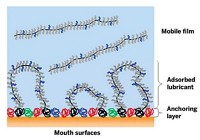
In natural body joints, surfaces in sliding contact are lubricated and protected by a thin film of synovial fluid, which is a complex mixture of proteins, hyaluronan, phospholipids, and other compounds. To begin to understand the roles played by those components in metal-on-metal hips, Cann and her Imperial College coworker Connor W. Myant conducted laboratory simulations that mimicked the variety of stop-and-go motions and varying forces experienced by real joints, for example when a person stands, begins walking, stops, and sits.

The team used a series of films of bovine calf serum solutions as stand-ins for synovial fluid and measured various changes in the films as they were compressed between a Co-Cr-Mo surface sliding against a glass-disk reference surface. The tests indicate that the films undergo changes in thickness and flow properties as a result of tribology-driven protein aggregation in the synovial fluid entrained in the contact region. That picture of lubricant dynamics, which differs from conventional ones, could be used to modify prosthesis geometry to extend device lifetimes, the team suggests (J. Mech. Behav. Biomed. Mater. 2014, DOI: 10.1016/j.jmbbm.2013.12.016).
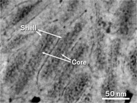
An electron microscopy study revealed another type of tribology-induced change in these lubricating films—an elemental one. A couple of years ago, Laurence D. Marks of Northwestern University, Markus A. Wimmer of Rush University Medical Center, and coworkers examined several metal-on-metal hip implants that were removed from patients. The group’s aim was to characterize the films that formed inside the body between implant surfaces in sliding contact. The team unexpectedly found a nanometer-thick film of graphitic carbon—a surface-protecting solid lubricant that was not present in unused implants. The graphite-forming mechanism has not yet been determined but may involve tribochemical conversion of lubricant proteins.
In a follow-up study, the team, which includes Rush University’s Michel P. Laurent, simulated the film-forming process by rubbing a reference ceramic material against a Co-Cr-Mo surface in the presence of a synovial fluid mimic. The experiment was carried out in a tribometer designed to measure various surface changes, including corrosion, which was monitored electrochemically. The tests show that the film protects the underlying surface from intense abrasive wear and corrosion. In addition, the film forms only when the rubbing force is comparable to forces in hip joints. At lower forces, the film does not form, and at higher forces, it decomposes.
Tribology isn’t always to blame for implant corrosion. According to Syracuse University’s Jeremy L. Gilbert, phagocytic (or inflammatory) cells do that job handily by generating reactive oxygen species. Gilbert and coworkers examined numerous hip and knee implants that had been removed from patients’ bodies. They found that regions of Co-Cr-Mo surfaces that were not in sliding contact with other surfaces often exhibited telltale marks of cell-induced corrosion. The surfaces were patterned with cellular remains and biological materials, showing evidence of attacks in which cells attached, spread, and migrated across implant surfaces (J. Biomed. Mater. Res., Part A 2014, DOI: 10.1002/jbm.a.35165).
Fifteen years have passed since Medley’s follow-up hip replacement surgery in which an old-style polyethylene component was replaced with an improved one. This time things look good. “I haven’t had any trouble with it yet,” he says. But the seasoned tribologist is practical, adding, “I may have to have another revision at some stage.”
Ongoing improvements in joint implants and deeper understanding of wear-and-tear processes may afford next-generation patients a far bolder outlook. For them, joint-implant surgery may be a case of one and done.


Join the conversation
Contact the reporter
Submit a Letter to the Editor for publication
Engage with us on Twitter