Advertisement
Grab your lab coat. Let's get started
Welcome!
Welcome!
Create an account below to get 6 C&EN articles per month, receive newsletters and more - all free.
It seems this is your first time logging in online. Please enter the following information to continue.
As an ACS member you automatically get access to this site. All we need is few more details to create your reading experience.
Not you? Sign in with a different account.
Not you? Sign in with a different account.
ERROR 1
ERROR 1
ERROR 2
ERROR 2
ERROR 2
ERROR 2
ERROR 2
Password and Confirm password must match.
If you have an ACS member number, please enter it here so we can link this account to your membership. (optional)
ERROR 2
ACS values your privacy. By submitting your information, you are gaining access to C&EN and subscribing to our weekly newsletter. We use the information you provide to make your reading experience better, and we will never sell your data to third party members.
Analytical Chemistry
Dye Colors Up Live Cell Surfaces In 3-D
Photoswitchable dye covalently labels amines on bacteria surfaces to help create superresolution three-dimensional images
by Celia Henry Arnaud
October 6, 2014
| A version of this story appeared in
Volume 92, Issue 40

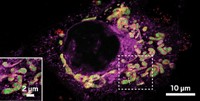
Many organic dyes otherwise suitable for superresolution microscopy require ultraviolet light to turn on their fluorescence, which can damage live cells. A new rhodamine spirolactam, developed by W. E. Moerner of Stanford University, Robert J. Twieg of Kent State University, and coworkers, is more compatible with live-cell imaging because it switches from its nonfluorescent form to its fluorescent isomer when irradiated with visible light. The researchers have used the dye to acquire three-dimensional images of commonly studied Caulobacter crescentus bacteria cells (J. Am. Chem. Soc. 2014, DOI: 10.1021/ja508028h). The positively charged dye, which doesn’t cross the bacterial cell membrane, contains a N-hydroxysuccinimide ester that covalently labels amines on the cell surface. The researchers turn on the fluorescence of a few dye molecules at a time with a low-intensity 405-nm purple laser and read the fluorescence with a 561-nm green laser. By doing this many times, they build superresolution images of the live cell surfaces in which they are able to resolve stalk structures that aren’t visible using standard microscopy.


Join the conversation
Contact the reporter
Submit a Letter to the Editor for publication
Engage with us on Twitter