Advertisement
Grab your lab coat. Let's get started
Welcome!
Welcome!
Create an account below to get 6 C&EN articles per month, receive newsletters and more - all free.
It seems this is your first time logging in online. Please enter the following information to continue.
As an ACS member you automatically get access to this site. All we need is few more details to create your reading experience.
Not you? Sign in with a different account.
Not you? Sign in with a different account.
ERROR 1
ERROR 1
ERROR 2
ERROR 2
ERROR 2
ERROR 2
ERROR 2
Password and Confirm password must match.
If you have an ACS member number, please enter it here so we can link this account to your membership. (optional)
ERROR 2
ACS values your privacy. By submitting your information, you are gaining access to C&EN and subscribing to our weekly newsletter. We use the information you provide to make your reading experience better, and we will never sell your data to third party members.
Analytical Chemistry
Imaging dangerous dendrite growth inside a Li-ion battery
MRI method with high spatial and temporal resolution provides close-up look at uncontrolled metal accumulation
by Mitch Jacoby
September 26, 2016
| A version of this story appeared in
Volume 94, Issue 38
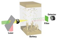
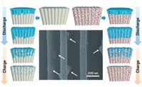
By devising a method that provides a detailed view inside a lithium-ion battery while it is running, researchers have come up with a way to gather reconnaissance information about processes that could trigger disastrous battery failure. Under some charging conditions, lithium can accumulate on a battery’s anode, leading to uncontrolled growth of needlelike metal dendrites that can cause hazardous short circuiting. Lithium metal, an ideal anode material based on its exceptional charge-storage capacity, carries a high dendrite risk. So manufacturers use lower capacity carbon anodes, which are safer but not fully dendrite-proof. To study this poorly understood process, Andrew J. Ilott and Alexej Jerschow of New York University and coworkers developed an 1H magnetic resonance imaging (MRI) method and used it to watch dendrites grow inside a Li-ion battery as it was being charged (Proc. Nat. Acad. Sci. USA 2016, DOI: 10.1073/pnas.1607903113). Unlike an earlier Li MRI method developed by the team, which images dendrites directly, the new method measures “shadows” formed by the magnetic properties of the growing dendrites in the surrounding electrolyte solution. Because the shadows are roughly 20 times as large as the dendrites, the new method provides much improved spatial resolution. It also boosts the time resolution, owing to the relatively shorter scan times for 1H NMR.


Join the conversation
Contact the reporter
Submit a Letter to the Editor for publication
Engage with us on Twitter