Advertisement
Grab your lab coat. Let's get started
Welcome!
Welcome!
Create an account below to get 6 C&EN articles per month, receive newsletters and more - all free.
It seems this is your first time logging in online. Please enter the following information to continue.
As an ACS member you automatically get access to this site. All we need is few more details to create your reading experience.
Not you? Sign in with a different account.
Not you? Sign in with a different account.
ERROR 1
ERROR 1
ERROR 2
ERROR 2
ERROR 2
ERROR 2
ERROR 2
Password and Confirm password must match.
If you have an ACS member number, please enter it here so we can link this account to your membership. (optional)
ERROR 2
ACS values your privacy. By submitting your information, you are gaining access to C&EN and subscribing to our weekly newsletter. We use the information you provide to make your reading experience better, and we will never sell your data to third party members.
Analytical Chemistry
Revealing 3-D organization of meiosis complex
Combining two kinds of microscopy reveals a layered structure in the synaptonemal complex
by Celia Henry Arnaud
August 7, 2017
| A version of this story appeared in
Volume 95, Issue 32

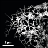
A multiprotein structure called the synaptonemal complex helps chromosomes segregate properly during the cell division process known as meiosis. Although much is already known about the proteins in the complex and their two-dimensional arrangement, the 3-D organization of the complex is less well understood. Now, a team led by R. Scott Hawley, Brian D. Slaughter, and Jay R. Unruh of Stowers Institute for Medical Research has combined two microscopy techniques to get a better 3-D picture of the fruit fly version of the complex (Proc. Natl. Acad. Sci. USA 2017, DOI: 10.1073/pnas.1705623114). The researchers modified a technique called expansion microscopy to make it compatible with superresolution microscopy. In expansion microscopy, a gel matrix with an embedded sample is uniformly inflated, which separates the components in the sample while keeping them correctly oriented relative to one another. To use the method with superresolution microscopy, the researchers modified the method so they could thinly slice the samples after embedding them in the matrix but before expanding the matrix. They expanded the samples to about four times their normal size. Then they acquired images using a superresolution microscopy method called structured illumination microscopy. The images revealed that the complex has two layers that mirror each other. The researchers propose that the layers possibly connect a sister chromatid (one of two identical copies of a replicated chromosome) from one homologous chromosome to a sister chromatid of the other homolog.


Join the conversation
Contact the reporter
Submit a Letter to the Editor for publication
Engage with us on Twitter