Advertisement
Grab your lab coat. Let's get started
Welcome!
Welcome!
Create an account below to get 6 C&EN articles per month, receive newsletters and more - all free.
It seems this is your first time logging in online. Please enter the following information to continue.
As an ACS member you automatically get access to this site. All we need is few more details to create your reading experience.
Not you? Sign in with a different account.
Not you? Sign in with a different account.
ERROR 1
ERROR 1
ERROR 2
ERROR 2
ERROR 2
ERROR 2
ERROR 2
Password and Confirm password must match.
If you have an ACS member number, please enter it here so we can link this account to your membership. (optional)
ERROR 2
ACS values your privacy. By submitting your information, you are gaining access to C&EN and subscribing to our weekly newsletter. We use the information you provide to make your reading experience better, and we will never sell your data to third party members.
Spectroscopy
Raman spectroscopy, now with Angstrom-level resolution
Imaging study yields atomic-resolution images of molecular vibrations
by Mitch Jacoby
April 11, 2019
| A version of this story appeared in
Volume 97, Issue 15

By combining scanning probe microscopy, laser spectroscopy, and surface science, researchers have sharpened the spatial resolution of microspectroscopy to the angstrom level. The study demonstrates a way to record vibrational spectra from select regions within a single molecule and a technique for capturing images of that molecule’s modes of vibration (Nature 2019, DOI: 10.1038/s41586-019-1059-9).
Raman spectroscopy is a powerful tool for probing molecular vibrations. It’s used commercially for quality and process control and in research settings to study molecular phenomena. But the Raman signal, produced by sample molecules interacting with light—often visible light from lasers—is weak, limiting the method’s sensitivity.
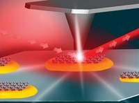
Over 40 years ago, researchers discovered that Raman signals could be boosted by a factor greater than one million by adsorbing analyte molecules on rough metal surfaces, such as silver, patterned with microscopically sharp points. The metal spikes acted like antennae, amplifying the electromagnetic field in nanometer-sized regions and enhancing the signal.

Finding those nanosized hot spots and getting molecules to adsorb there is tricky. Researchers soon gained more control by using ultrasharp metal tips in scanning probe microscopy to carefully bring the antennae to the molecules. With tip-enhanced Raman spectroscopy (TERS), scientists can record Raman spectra from individual molecules within a few nanometers of the tip.
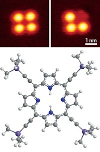
In the new study, Joonhee Lee and V. Ara Apkarian of the University of California, Irvine, and coworkers go a step further. Using cobalt tetraphenyl porphyrin as a test case, the team demonstrated that TERS can be used to record unique spectra from various regions within a single molecule. The spectra differ with angstrom resolution as the team moves the tip from one spot, for example over a phenyl ring—exciting a specific C–H stretch—to another part of the molecule.
“This is really elegant work,” says Renee Frontiera, a laser spectroscopist at the University of Minnesota, Minneapolis. The specific ways a molecule vibrates are typically inferred from theory, due to limits in imaging resolution. But Apkarian’s group has come up with a way to map the molecule’s vibrational modes with unprecedented spatial resolution. She adds, “with this work, it’s now possible to see how energy deposited in one specific region of the molecule flows throughout the whole structure.”
Eric C. Le Ru, a specialist in nanoscale optical phenomena, is also impressed with the study. He notes, however, that the specialized vacuum equipment and cryogenic temperatures used here may limit the method’s applicability. Also, the success depended on the porphyrin interacting strongly with a copper surface on which it was anchored. Other combinations of molecules and surfaces may not lead to the same resolution.
Frontiera agrees. Even so, she says “I hope these images show up in the vibrational spectroscopy chapter of future chemistry textbooks.”
CORRECTION:
This story was updated on April 17, 2019, to correct the structure for cobalt tetraphenyl porphyrin. Because of a production error, it appeared without charges.


Join the conversation
Contact the reporter
Submit a Letter to the Editor for publication
Engage with us on Twitter