Advertisement
Grab your lab coat. Let's get started
Welcome!
Welcome!
Create an account below to get 6 C&EN articles per month, receive newsletters and more - all free.
It seems this is your first time logging in online. Please enter the following information to continue.
As an ACS member you automatically get access to this site. All we need is few more details to create your reading experience.
Not you? Sign in with a different account.
Not you? Sign in with a different account.
ERROR 1
ERROR 1
ERROR 2
ERROR 2
ERROR 2
ERROR 2
ERROR 2
Password and Confirm password must match.
If you have an ACS member number, please enter it here so we can link this account to your membership. (optional)
ERROR 2
ACS values your privacy. By submitting your information, you are gaining access to C&EN and subscribing to our weekly newsletter. We use the information you provide to make your reading experience better, and we will never sell your data to third party members.
Analytical Chemistry
Taking Biomolecules for a Spin
Electron spin resonance answers questions that can't be answered with NMR or crystallography
by CELIA M. HENRY, C&EN WASHINGTON
March 28, 2005
| A version of this story appeared in
Volume 83, Issue 13
Electron spin resonance answers questions that can't be answered with NMR or crystallography
The answers to many biological questions require structural information about molecules such as proteins and nucleic acids. People usually turn to X-ray crystallography or nuclear magnetic resonance spectroscopy to acquire these structures. When those methods don't work, electron paramagnetic resonance spectroscopy can help.
EPR (also called electron spin resonance spectroscopy) complements crystallography and NMR for proteins that don't crystallize or are too big for NMR. In EPR, an unpaired electron absorbs microwave radiation in the presence of a strong magnetic field. The resulting change in the electron's spin states reveals information about the structure and dynamics of the molecule.
EPR is especially useful for integral membrane proteins, which don't crystallize easily and can be larger than 200 kilodaltons. Yet "10 or 15 years ago, people thought that EPR would never work on integral membrane proteins" because the proteins don't have an unpaired electron, according to Sunil Saxena, an assistant professor of chemistry at the University of Pittsburgh and the organizer of a symposium on EPR at the Pittsburgh Conference, held earlier this month in Orlando, Fla. Biochemists have used EPR to look at metalloproteins because they contain metal centers that could harbor unpaired electrons, but the information obtained is localized around the metal center, he explained.
Things changed after Wayne L. Hubbell's group at the University of California, Los Angeles, developed a method for introducing spin labels anywhere in transmembrane proteins: Attach a nitroxide to the protein at a cysteine residue, which can be added by replacing the natural amino acid with a cysteine through site-directed mutagenesis. With labels installed in the protein, EPR yields a spectrum that furnishes information about the protein's secondary structure and, if it's a membrane protein, how deeply the label is buried in the membrane.
If proteins are tagged with two spin labels, the magnetic dipole interaction between the labels creates a distance-dependent splitting in the EPR spectrum, from which the distance between the labels can be calculated. Doing this for spin labels on different pairs of amino acids generates a set of distance constraints that can be used to generate a structure. Unfortunately, compared with the broad pattern of the spectrum, the splitting caused by the dipole interaction is minuscule, Saxena said.
To obtain a sharper spectrum, instead of measuring one spin flip, Saxena and his coworkers look at two spin flips in a method called double-quantum Fourier transform EPR, in which the microwave radiation is pulsed. The experiment yields a spectrum that is better resolved than that from normal one-dimensional EPR.
Saxena developed the initial methodology when he was a graduate student with Jack H. Freed at Cornell University. At Pittsburgh, "when I started this double-quantum work three years ago, there was genuine concern that it would not work on proteins, let alone large membrane proteins," Saxena told C&EN.

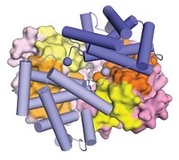
HE'S PROVING the naysayers wrong. Saxena, in collaboration with Michael Cascio, professor of molecular genetics and biochemistry at Pittsburgh, is using the double-quantum method to generate structural information about the glycine receptor, an approximately 250-kDa membrane protein involved in cell signaling and communication. It consists of five identical subunits that form a pore that, when opened by glycine binding, allows chlorine to pass through. Right now, biologists have a "cartoon understanding" of the receptor, Saxena said.
To obtain structural information, Saxena's team labels the same amino acid on each of the subunits. Because the subunits are symmetrically arranged, the five labels correspond to only two unique distances. By labeling cysteines at different points in the subunits, Saxena has found that the subunits are closer to each other in the portion that is buried in the membrane than they are in the region outside the membrane: The protein is funnel shaped.
Beyond seeking structural information, researchers are now using EPR to answer all sorts of biological questions, ranging from the folding of prions to the formation of protein fibrils to how membrane fusion occurs.
Madhuri Chattopadhyay, a graduate student with Glenn L. Millhauser in the chemistry department at the University of California, Santa Cruz, described the group's work to understand the structure and function of the prion protein.
The diseases associated with the prion protein are caused by its misfolding, but scientists still don't know the function of the normal prion. It has been implicated in the regulation of apoptosis, protection from oxidative stress, and copper transport. A region of repeating eight-amino acid sequences, which bind Cu(II), is involved with each of these functions.
Using EPR, Millhauser's group is trying to understand the protein's binding to copper (Acc. Chem. Res. 2004, 37, 79). The EPR spectrum suggests two modes of binding. One dominates when the protein is fully loaded with copper. The other is seen at low pH and when the protein is only partially loaded with Cu(II).
To find out the minimum sequence needed to bind a single Cu(II), they designed a peptide library based on the octarepeat, including peptides with different frequencies of the repeated sequence and peptides with shortened versions of the octarepeat. Using several EPR techniques, including multifrequency and pulsed EPR, they found that a five-amino acid region consisting of a histidine, three glycines, and a tryptophan was the minimum component required to bind Cu(II). They believe that the binding involves deprotonated amide nitrogen atoms and amide carbonyl oxygen atoms.
The binding site shows up four times in the prion protein, according to the EPR spectrum. When Millhauser and coworkers titrated the protein for copper binding, however, they found that five or even six copper ions can bind to the protein. They propose that the other binding site--which consists of glycine, threonine, and histidine--is in a different region of the protein and that it involves a binding mode that is different from that in other sites.
EPR is being used to look at other disease-related proteins as well. For example, Ralf Langen, an assistant professor of biochemistry and molecular biology at the University of Southern California, is studying amyloid protein structures. Such proteins play a role in a number of diseases, including Alzheimer's disease, Parkinson's disease, and type 2 diabetes.
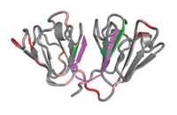
Langen uses EPR to study the structures of the fibrils, which are aggregates of individual filament proteins and the structures of which are poorly understood. All known amyloid fibrils contain β-strands, running perpendicular to the fiber axis. Whether those β-strands are arranged parallel or antiparallel to one another is largely unknown, as is the three-dimensional structure of fibrils.
At the symposium, Langen described work on the tau protein, which is associated with Alzheimer's disease and dementia. The tau monomers are believed to be random coils. Heparin and other anionic cofactors greatly accelerate the formation of filaments from the monomers.
Line-shape analysis of the EPR spectrum indicates a tight packing of the filaments formed by tau. In addition, Langen found that tau consists of core residues in its C-terminal half that stack on top of each other along the fiber axis (Proc. Natl. Acad. Sci. USA 2004, 101, 10278). Similar stacking is observed in β-helices in which strands wind around one another to form multiple layers. In the case of tau filaments, however, the monomers stack on top of each other, forming single layers.
Meanwhile, Yeon-Kyun Shin, a professor of biochemistry, biophysics, and molecular biology at Iowa State University, is using EPR to study the fusion of plasma and vesicle membranes mediated by a protein complex called SNARE. The complex consists of proteins known as VAMP, SNAP-25, and syntaxin. Using EPR, Shin has shown that SNARE-mediated membrane fusion proceeds through a process called hemifusion, in which the inner layers of the membrane bilayers fuse first.

ALTHOUGH MUCH of the work in EPR involves proteins, the method isn't just for proteins anymore. For example, Snorri T. Sigurdsson, a chemistry professor at the University of Iceland, in Reykjavk, has used EPR to study the interactions between RNA and its ligands. General techniques for spin labeling nucleic acids have only recently become available, he said. Thus, commercially available RNAs containing 2'-amino pyrimidine nucleotides at specific sites can be readily spin labeled by reacting the 2' amino group with an isocyanate spin-labeling reagent. When ligands bind to the labeled RNA, they change the EPR spectrum.
Sigurdsson has been studying the TAR RNA, which is involved in HIV replication by interacting with the Tat protein during transcription. Both X-ray and NMR structures of the TAR RNA are available, but there are differences between the crystal and solution-state structures; these differences have been suggested to be due to calcium binding in the crystallized form.

To verify whether the structural differences are specific to calcium binding, Sigurdsson analyzed dynamic signatures. In this technique, the change in spectral width of the EPR spectrum, which reflects a change in mobility, is plotted as a function of spin-label position in a bar graph for several labeled nucleotides. Both the magnitude and the direction of the change are important components of the signature. He has found that the nucleic acid produces the same signature when interacting with calcium or sodium. That result indicates that the structural differences are not specific to calcium.
EPR dynamic signatures could be used to assess the binding mode of small-molecule ligands from a collection of compounds known to bind the TAR RNA, Sigurdsson said. "Everything we did confirmed that compounds that bind in different ways give different dynamic signatures, whereas compounds that bind in a similar manner have nearly identical signatures." For targeting RNA with drugs, EPR measurements could be used in the discovery efforts to identify the binding site of different drug candidates, he suggested.
Sigurdsson also has used dynamic signatures to study the specific binding of TAR RNA to the Tat protein. He collected EPR signatures for a number of mutant Tat-derived peptides to determine which components of the protein are important for binding. Specific arginine residues on the C-terminal side of the essential arginine-52 turn out to be particularly important for Tat binding. "These results show that one can learn about protein structural determinants by studying the internal dynamics of the RNA," he said.
One of Saxena's goals for the symposium was to let people know that EPR has come a long way. "EPR now provides a very robust and powerful methodology for handling problems that cannot be routinely handled by NMR and X-ray," he said.


Join the conversation
Contact the reporter
Submit a Letter to the Editor for publication
Engage with us on Twitter