Advertisement
Grab your lab coat. Let's get started
Welcome!
Welcome!
Create an account below to get 6 C&EN articles per month, receive newsletters and more - all free.
It seems this is your first time logging in online. Please enter the following information to continue.
As an ACS member you automatically get access to this site. All we need is few more details to create your reading experience.
Not you? Sign in with a different account.
Not you? Sign in with a different account.
ERROR 1
ERROR 1
ERROR 2
ERROR 2
ERROR 2
ERROR 2
ERROR 2
Password and Confirm password must match.
If you have an ACS member number, please enter it here so we can link this account to your membership. (optional)
ERROR 2
ACS values your privacy. By submitting your information, you are gaining access to C&EN and subscribing to our weekly newsletter. We use the information you provide to make your reading experience better, and we will never sell your data to third party members.
Materials
Imaging Tumors With Degradable Nanoparticles
Fluorescent, porous silicon particles can also carry drugs in vivo
by Rachel Petkewich
February 24, 2009

The first fluorescent nanoparticles that can circulate in the blood and degrade into nontoxic by-products could also help image tumors and carry cancer-fighting drugs. Michael J. Sailor, a chemist at the University of California, San Diego, and colleagues developed the porous silicon nanoparticles (Nature Mater., DOI: 10.1038/nmat2398).
Although other fluorescent nanoparticles such as quantum dots are available for imaging studies, he says, the brightest dots contain cadmium selenide, which is too toxic for use in humans, whereas silicon is actually a trace nutrient in the body. In addition, porous silicon can carry drugs, unlike most nanoparticles, which are solid.
The idea is to use the inherent luminescence of the particles to track them as they chaperone the drug directly to a tumor rather than to other parts of the body and then let them degrade harmlessly, Sailor adds.
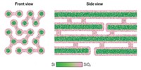
Sailor and his colleagues used electrochemical machining techniques to create porous silicon. They applied electrical currents to silicon wafers in an ethanolic hydrogen fluoride solution and used ultrasonic waves to break the porous wafers into 126-nm-diameter particles with 5- to 10–nm-diameter pores. Chemically degrading the surfaces of the silicon particles yielded a silicon dioxide layer that is luminescent under ultraviolet light. In mice, such particles hydrolyze to silicic acid by-products that are eliminated via urine in about 24 hours.
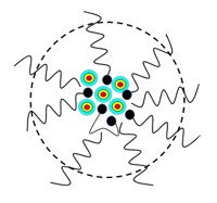
The researchers loaded these degradable particles with the anticancer drug doxorubicin. They injected the particles into a mouse with a tumor and demonstrated with imaging that the particles aggregate in the tumor. To ensure that the particles could circulate in the body long enough to be useful, Sailor's team coated them with the polysaccharide dextran.
Jarno Salonen, a physicist who studies drug delivery applications of porous silicon at the University of Turku, in Finland, praised this "pioneering" development. By modifying the pore size or the surface chemistry, one could tweak, for example, the particles' biodegradation rate as necessary, he says. He adds that porous silicon is quite versatile and can also be biofunctionalized if needed.


Join the conversation
Contact the reporter
Submit a Letter to the Editor for publication
Engage with us on Twitter