Advertisement
Grab your lab coat. Let's get started
Welcome!
Welcome!
Create an account below to get 6 C&EN articles per month, receive newsletters and more - all free.
It seems this is your first time logging in online. Please enter the following information to continue.
As an ACS member you automatically get access to this site. All we need is few more details to create your reading experience.
Not you? Sign in with a different account.
Not you? Sign in with a different account.
ERROR 1
ERROR 1
ERROR 2
ERROR 2
ERROR 2
ERROR 2
ERROR 2
Password and Confirm password must match.
If you have an ACS member number, please enter it here so we can link this account to your membership. (optional)
ERROR 2
ACS values your privacy. By submitting your information, you are gaining access to C&EN and subscribing to our weekly newsletter. We use the information you provide to make your reading experience better, and we will never sell your data to third party members.
Analytical Chemistry
Technique Dives Into Cells
Microscope offers best three-dimensional resolution yet
by Celia Henry Arnaud
February 5, 2009

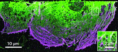
A new microscope improves the three-dimensional resolution of fluorescence imaging to an unprecedented level of detail. The instrument's resolution is better than 20 nm, at least twice as good as resolutions previously reported for 3-D fluorescence imaging.
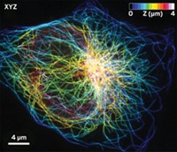
Harald F. Hess of Howard Hughes Medical Institute's Janelia Farm Research Campus, Jennifer Lippincott-Schwartz of the National Institutes of Health, and coworkers use their new method, called interferometric photoactivated localization microscopy (iPALM), to resolve such structures as microtubules, cell membranes, and receptors in the endoplasmic reticulum, the cellular organelle where protein and lipid synthesis occurs.
This is the first time microtubules, components of cells' cytoskeletons that are about 25 nm in diameter, have been resolved by a method other than electron microscopy, the researchers say. Furthermore, iPALM provides molecular information that is not available from electron microscopy.
The method can "dissect the molecular distribution of cellular machines that are nanoscopic in size," Lippincott-Schwartz says. "We're interested in looking at the 3-D characteristics of things that have only been well characterized by electron microscopy."
The new microscopy method combines PALM, a superresolution microscopy method developed previously by the same group, with single-photon multiphase interferometry (Proc. Natl. Acad. Sci. USA, DOI: 10.1073/pnas.0813131106). The key technology in the interferometric method is a newly developed three-way beam splitter that projects the photon beam onto three cameras. The interferometry technique adds a third dimension to the microscopy method by determining the vertical positions of fluorescent proteins from intensity ratios of the photon beam on the cameras.
So far, the researchers have performed iPALM with one fluorescent color at a time, but they plan to extend the method to accommodate multiple colors by developing an interferometric beam splitter that works over a wider wavelength range, Hess says.
"The volume resolvable by iPALM is about a factor of five to 10 better than any other optical 3-D microscopy technique so far," says microscopy expert Joerg Bewersdorf of Jackson Laboratory, in Bar Harbor, Maine. "This work therefore represents a big step toward true molecular resolution."


Join the conversation
Contact the reporter
Submit a Letter to the Editor for publication
Engage with us on Twitter