Advertisement
Grab your lab coat. Let's get started
Welcome!
Welcome!
Create an account below to get 6 C&EN articles per month, receive newsletters and more - all free.
It seems this is your first time logging in online. Please enter the following information to continue.
As an ACS member you automatically get access to this site. All we need is few more details to create your reading experience.
Not you? Sign in with a different account.
Not you? Sign in with a different account.
ERROR 1
ERROR 1
ERROR 2
ERROR 2
ERROR 2
ERROR 2
ERROR 2
Password and Confirm password must match.
If you have an ACS member number, please enter it here so we can link this account to your membership. (optional)
ERROR 2
ACS values your privacy. By submitting your information, you are gaining access to C&EN and subscribing to our weekly newsletter. We use the information you provide to make your reading experience better, and we will never sell your data to third party members.
Analytical Chemistry
A View Inside Tissues And Cells
by Lauren K. Wolf
March 8, 2010
| A version of this story appeared in
Volume 88, Issue 10

Ask X. Sunney Xie to identify ultrafast lasers' greatest impact on the life sciences, and his response is reflexive. "Imaging," he says, without skipping a beat. "Look at how many ultrafast lasers are made and sold for that purpose," says Xie, a chemistry professor at Harvard University and pioneer of CARS (coherent anti-Stokes Raman scattering) microscopy. "I think in recent years, it is clear the dominant market is for imaging."

Although the idea for confocal microscopy, a method that can acquire in-focus images of samples at select depths, was patented in the late 1950s, it took the invention of the laser to make the technique routine. Confocal laser scanning microscopes are now standard for three-dimensional imaging of cells, "particularly live cells," says Stefan W. Hell, a laser microscopy expert at the Max Planck Institute for Biophysical Chemistry, in Göttingen, Germany.
And in the past two decades, improvements in optical technology and the development of robust ultrafast lasers have caused an explosion of new and innovative microscopy techniques.
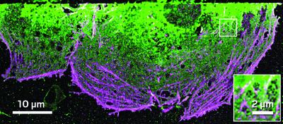
Superresolution fluorescence methods enable the imaging of fluorescently labeled biological structures with dimensions on the order of tens of nanometers. For instance, STED (stimulated emission depletion) microscopy, a technique pioneered by Hell, has made it possible to go beyond the diffraction-limited resolution of light. With it, his group has been able to look inside brain tissue slices at "the fine details in a living neuron," he says. "For many bioimaging applications," he adds, "scientists no longer have to resort to electron microscopy," a destructive method generally incapable of live-cell imaging.
Laser-based multiphoton methods, on the other hand, can't quite achieve this resolution, but some of them—CARS and stimulated Raman scattering microscopy, for instance—instead boast the ability to operate fluorophore-free. These techniques probe vibrational modes, such as C–H stretching motions, in molecules and have been used to image ultrafast dynamics in systems including the lipid domains in cell membranes and lipid metabolism in liver cells.
Although CARS and other laser microscopy methods don't have the tissue penetration depth of magnetic resonance imaging (MRI), the time and spatial resolution are much higher, Xie says. For this reason, he is working with collaborators to bring laser microscopy into hospital rooms and deeper into the human body by developing a CARS-compatible endoscope for cancer biopsies.
Similarly, Watt W. Webb, an applied physics professor at Cornell University, is working with collaborators to develop an endoscope for two-photon fluorescence microscopy, a technique he coinvented in 1990. Making use of intrinsic tissue fluorescence, Webb hopes to use the method for earlier detection of hard-to-diagnose conditions such as lung and bladder cancers. "There is no endoscope yet built that is geared up to work with multiphoton spectroscopy," he says. "That's where the frontier in imaging is."
Advertisement
You might also like...

The power is now in your (nitrile gloved) hands
Sign up for a free account to get more articles. Or choose the ACS option that’s right for you.
Already have an ACS ID? Log in
Join the conversation
Contact the reporter
Submit a Letter to the Editor for publication
Engage with us on Twitter