Advertisement
Grab your lab coat. Let's get started
Welcome!
Welcome!
Create an account below to get 6 C&EN articles per month, receive newsletters and more - all free.
It seems this is your first time logging in online. Please enter the following information to continue.
As an ACS member you automatically get access to this site. All we need is few more details to create your reading experience.
Not you? Sign in with a different account.
Not you? Sign in with a different account.
ERROR 1
ERROR 1
ERROR 2
ERROR 2
ERROR 2
ERROR 2
ERROR 2
Password and Confirm password must match.
If you have an ACS member number, please enter it here so we can link this account to your membership. (optional)
ERROR 2
ACS values your privacy. By submitting your information, you are gaining access to C&EN and subscribing to our weekly newsletter. We use the information you provide to make your reading experience better, and we will never sell your data to third party members.
Analytical Chemistry
A Clearer Picture Of Cell Secretion
Scientists combine electrochemical and optical measurements to study exocytosis
by Celia Henry Arnaud
May 9, 2011
| A version of this story appeared in
Volume 89, Issue 19
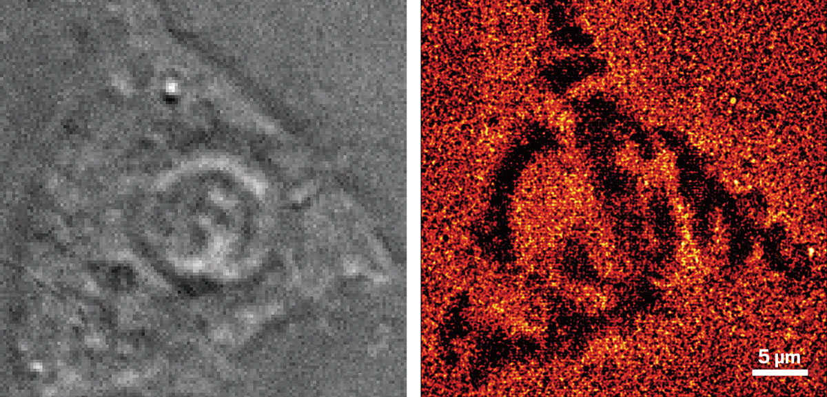
Combining electrochemical and optical measurements will give scientists a better window into exocytosis, a process by which cells secrete chemicals such as neurotransmitters, French researchers report (Angew. Chem. Int. Ed., DOI: 10.1002/anie.201101148). Electrochemical measurements have good time resolution, but they can’t detect early stages of exocytosis before vesicles fuse with the cell membrane and secrete their chemical cargo. In contrast, optical measurements can track individual vesicles before fusion begins but have poorer time resolution. Christian Amatore and coworkers at the École Normale Supérieure, in Paris, used transparent indium tin oxide electrodes to monitor the release of the neurotransmitter serotonin from cultured endocrine cells with amperometry; they used total internal reflection fluorescence (TIRF) microscopy of fluorescently labeled neuropeptide Y to visualize the vesicles. The 150-nm-thick band-shaped electrodes allowed sufficient resolution for TIRF without compromising the quality of the electrochemical measurements. The electrochemical and TIRF signals are obtained simultaneously, which allows information from the two methods to be correlated.
You might also like...


The power is now in your (nitrile gloved) hands
Sign up for a free account to get more articles. Or choose the ACS option that’s right for you.
Already have an ACS ID? Log in
Join the conversation
Contact the reporter
Submit a Letter to the Editor for publication
Engage with us on Twitter