Advertisement
Grab your lab coat. Let's get started
Welcome!
Welcome!
Create an account below to get 6 C&EN articles per month, receive newsletters and more - all free.
It seems this is your first time logging in online. Please enter the following information to continue.
As an ACS member you automatically get access to this site. All we need is few more details to create your reading experience.
Not you? Sign in with a different account.
Not you? Sign in with a different account.
ERROR 1
ERROR 1
ERROR 2
ERROR 2
ERROR 2
ERROR 2
ERROR 2
Password and Confirm password must match.
If you have an ACS member number, please enter it here so we can link this account to your membership. (optional)
ERROR 2
ACS values your privacy. By submitting your information, you are gaining access to C&EN and subscribing to our weekly newsletter. We use the information you provide to make your reading experience better, and we will never sell your data to third party members.
Biological Chemistry
Ca2+ Indicators Show Their New Colors
by Lauren K. Wolf
September 12, 2011
| A version of this story appeared in
Volume 89, Issue 37

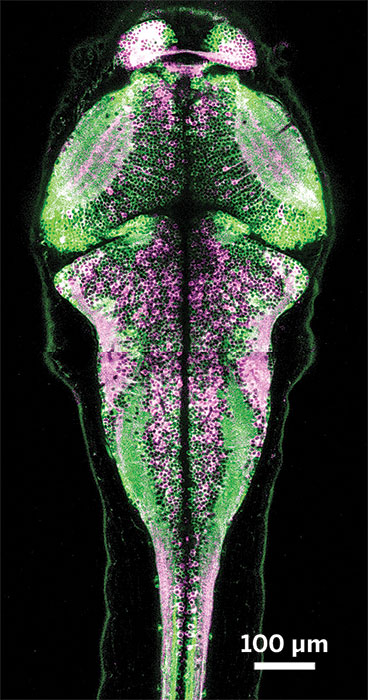
Previously containing a single, lonely splotch of green, the palette of genetically encoded calcium-ion indicators for fluorescent cellular imaging has now expanded to other colors, thanks to researchers from Japan and Canada (Science, DOI: 10.1126/science.1208592). These indicator proteins are important tools, especially in neuroscience, for measuring Ca2+ concentrations in cells. Starting with a set of green-light-emitting indicators called GCaMPs, the researchers, led by Robert E. Campbell of the University of Alberta, screened genetic variants of these proteins in Escherichia coli’s periplasm—the space between the microbe’s inner and outer membranes. The team developed red- and blue-emitting indicators, an improved green indicator, and a version that emits blue light when Ca2+ is bound and green light when it is not. These sensors, which the research team dubbed GECOs to “bring to mind the colorful family of lizards,” Campbell says, enable scientists to simultaneously image Ca2+ concentrations in multiple locations within the same cell. He adds that his team will soon release the indicator genes to a public repository “to get them into the hands of as many end users as possible.”
You might also like...


The power is now in your (nitrile gloved) hands
Sign up for a free account to get more articles. Or choose the ACS option that’s right for you.
Already have an ACS ID? Log in
Join the conversation
Contact the reporter
Submit a Letter to the Editor for publication
Engage with us on Twitter