Advertisement
Grab your lab coat. Let's get started
Welcome!
Welcome!
Create an account below to get 6 C&EN articles per month, receive newsletters and more - all free.
It seems this is your first time logging in online. Please enter the following information to continue.
As an ACS member you automatically get access to this site. All we need is few more details to create your reading experience.
Not you? Sign in with a different account.
Not you? Sign in with a different account.
ERROR 1
ERROR 1
ERROR 2
ERROR 2
ERROR 2
ERROR 2
ERROR 2
Password and Confirm password must match.
If you have an ACS member number, please enter it here so we can link this account to your membership. (optional)
ERROR 2
ACS values your privacy. By submitting your information, you are gaining access to C&EN and subscribing to our weekly newsletter. We use the information you provide to make your reading experience better, and we will never sell your data to third party members.
Biological Chemistry
Cells Keep Nanodisks Out
Nanoscience: Cells take up spherical nanoparticles but block entry by disk-shaped ones
by Alexander Hellemans
August 7, 2012

A team of Chinese and American researchers has demonstrated that disk-shaped nanoparticles bind to the cell membrane, but do not enter the interior of the cell (ACS Appl. Mater.Interfaces, DOI: 10.1021/am300840p). The information could help scientists design fluorescent nanoparticle tags that help track cells without harming them, the team says.
Researchers had previously found that nanoparticle size affected how the particles interacted with cells. A nanoparticle had to be a specific size—not too small or too large—to penetrate through a cell’s membrane (Adv. Mater., DOI: 10.1002/adma.200801393). A modeling study also suggested that the shape of carbon-based nanoparticles mattered: The cell membrane would block flat nanoparticles from entering, but let nanospheres in (J. Phys. Chem. B., DOI: 10.1021/jp0565148).
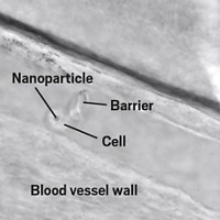
To find out experimentally whether the model was correct, Bing Yan of Shandong University, in China, and his colleagues compared how spherical and disk-shaped nanoparticles of the same diameter, 20 nm, interact with cells. They made nanodisks by polymerizing styrene, divinylbenzene, and butylstyrene, and bought commercial nanospheres. They added either nanodisks or nanospheres to HeLa cells and to other human cells. Using techniques including transmission electron microscopy imaging and fluorescence laser microscopy, they found that the nanodisks remain largely confined to the cell membrane, while the nanospheres cross the membrane into the cell’s interior.
The researchers think that the reason for the differing behavior is geometry: A nanodisk makes contact over a wider area with the cell membrane and sticks to it, while a spherical particle merely indents the membrane. As a result, nanospheres trigger endocytosis, a mechanism cells often use to pull in molecules, whereby the membrane engulfs the particle, explains Yan.
Because they do not venture inside cells, disk-shaped nanoparticles could play an important role in tracking stem cells, says Yan. “You want to label the cells but you don’t want to cause toxicity,” he says. Previous studies have found that when nanoparticles enter cells, they can kill the cells. The team found that, unlike nanospheres, nanodisks did not cause toxicity or even disturb the cell cycle The nanodisks also could help researchers study the cell membrane itself, says Yan, if the nanoparticles carried labels that attach to membrane receptors.
Giuseppe Battaglia of Sheffield University, in the U.K., says that the work illustrates the importance of nanoparticle shape in interactions with the cell. “Tubular nanoparticles also have a way of escaping uptake into the cell,” he says, though he suspects that they stick to membranes less effectively than Yan’s nanodisks do. Knowing how the various shapes interact with cells, he says, “is important information for people using nanoparticles for medical applications,” he says.


Join the conversation
Contact the reporter
Submit a Letter to the Editor for publication
Engage with us on Twitter