Advertisement
Grab your lab coat. Let's get started
Welcome!
Welcome!
Create an account below to get 6 C&EN articles per month, receive newsletters and more - all free.
It seems this is your first time logging in online. Please enter the following information to continue.
As an ACS member you automatically get access to this site. All we need is few more details to create your reading experience.
Not you? Sign in with a different account.
Not you? Sign in with a different account.
ERROR 1
ERROR 1
ERROR 2
ERROR 2
ERROR 2
ERROR 2
ERROR 2
Password and Confirm password must match.
If you have an ACS member number, please enter it here so we can link this account to your membership. (optional)
ERROR 2
ACS values your privacy. By submitting your information, you are gaining access to C&EN and subscribing to our weekly newsletter. We use the information you provide to make your reading experience better, and we will never sell your data to third party members.
Materials
Wiring Up Living Tissue
Sensing: Merging biology and electronics could lead to smart implants and prosthetics
by Bethany Halford
August 30, 2012

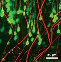
Science and science fiction moved a little closer together with a report that researchers have created so-called cyborg tissue that seamlessly integrates electronics with living cells (Nat. Mater., DOI: 10.1038/nmat3404). The new synthetic biomaterial could be used to probe how cells respond to drugs and might one day lead to smart prosthetics and tissue implants that sense and respond to their biological environment.
In recent years, researchers have developed several examples of flexible and stretchable electronics interfaced with tissue at its surface, explains Harvard University chemistry professor Charles M. Lieber, who spearheaded creation of the cyborg tissue along with Harvard Medical School’s Daniel S. Kohane. The new cyborg tissue takes the bioelectronics interface even further, Lieber says, by integrating the electronics within the tissue in a three-dimensional manner.
“This is the ultimate extension of the science fiction concept of a cyborg because we’re essentially merging electronic circuitry with biological circuitry,” Lieber says. “The electronics now look like the natural extracellular matrix.”
To intimately connect the electronic and biological components, Lieber and Kohane’s team struck upon the idea of creating a 3-D scaffold of biocompatible, nanoscale electronics that would provide mechanical support—just as the extracellular matrix does—for tissue to grow upon. They created a mesh of silicon wires, approximately 80 nm in diameter, upon which they constructed field-effect transistors at various points via lithography. The researchers then rolled the mesh or let it spring by design into a 3-D shape. Finally, they seeded the mesh with various types of cells, which they then coaxed to grow into tissue.
In one example, the team seeded the electronic scaffold with cardiac cells. They were able to detect the electrical signals the cells generated from within the engineered tissue in response to noradrenaline, a drug that stimulates cardiac contraction.
“There’s a whole toolbox of nanoelectronic devices that one can think about building into these 3-D circuits and merging them with biological information processing systems,” Lieber says, citing photonic devices as one example.
“This is really exciting work,” comments Yi Cui, a nanomaterials expert at Stanford University. “The concept of incorporating electronic sensors into a 3-D scaffold for local sensing in tissue opens up exciting opportunities. It can provide localized real-time monitoring of cellular activities and physicochemical change,” he says.
“This research opens a new direction in interfacing nanoelectronics networks with biological systems at an unprecedented level,” adds Zhong Lin Wang, a professor of nanoscience at Georgia Tech.


Join the conversation
Contact the reporter
Submit a Letter to the Editor for publication
Engage with us on Twitter