Advertisement
Grab your lab coat. Let's get started
Welcome!
Welcome!
Create an account below to get 6 C&EN articles per month, receive newsletters and more - all free.
It seems this is your first time logging in online. Please enter the following information to continue.
As an ACS member you automatically get access to this site. All we need is few more details to create your reading experience.
Not you? Sign in with a different account.
Not you? Sign in with a different account.
ERROR 1
ERROR 1
ERROR 2
ERROR 2
ERROR 2
ERROR 2
ERROR 2
Password and Confirm password must match.
If you have an ACS member number, please enter it here so we can link this account to your membership. (optional)
ERROR 2
ACS values your privacy. By submitting your information, you are gaining access to C&EN and subscribing to our weekly newsletter. We use the information you provide to make your reading experience better, and we will never sell your data to third party members.
Materials
Hearing Cells Poked By Tiny Probe
Nanotechnology: A nanoprobe simulates the effects of high-frequency sound waves on inner ear hair cells
by Erika Gebel
December 6, 2012

A nanosized probe can mimic sound waves by tickling microscopic hairlike projections on inner ear cells (Nano Lett., DOI: 10.1021/nl3036349). The device could help researchers understand how the cells turn sound into electrical signals the brain can interpret, the probe’s developers say.

Sound vibrations perturb bundles of “hair” that extend from cells in the cochlea of the inner ear. The perturbation causes ion channels in the cells’ membranes to open, creating electrical signals that are eventually relayed to the brain. “How that ion channel works isn’t known,” says Joey Doll, a former student in the laboratory of Beth Pruitt at Stanford University, who now works at a semiconductor company called SiTime.
To study the channels, scientists usually stimulate hair cell bundles with a glass rod about 10 μm across. Each hairlike fiber has a diameter of 500 nm and they bundle together in groups of 30 to 300 on each cell. These bundles respond to a wide range of frequencies of sound waves. But, because of the glass probe’s large size, it moves slowly and as a result, can mimic only low-frequency sounds, Doll says. Doll, Pruitt, and colleagues wanted to access higher frequencies so they built a smaller probe.
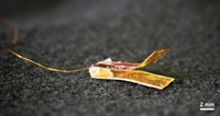
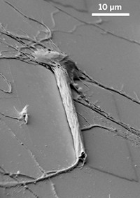
Their nanoprobe consists of two parts: a sensor and an actuator. The actuator generates the sensor’s motion, converting an electric current to a mechanical force. The sensor, just 2 μm wide, pushes on the hair cell bundles. When the researchers switch on the current, the probe tip flicks the hair cells to simulate sound vibrations.
To test the device, researchers removed ear tissue from rats and placed it under a microscope. Then, using a micromanipulator, they positioned the nanoprobe near a hair cell bundle and placed an electrode on the cell to detect the ion channel opening. For comparison, the researchers repeated the experiment with a glass probe. The nanoprobe moved far faster than the glass probe, achieving frequencies up to 80 kHz compared to the glass probe’s 10 kHz. Both devices triggered the opening of ion channels, according to patch clamp measurements.
Perturbing the cell at high frequencies hasn’t been done before this study, says Dolores Bozovic of the University of California, Los Angeles. She hopes the researchers will next turn their attention to the method that records the cells’ electrical signals, since their nanoprobe can stimulate the cells faster than they can measure changes in current. Indeed, Doll says the laboratory is seeking to do so while also exploring additional applications for the device such as studying the adhesive forces between bacteria in a biofilm.


Join the conversation
Contact the reporter
Submit a Letter to the Editor for publication
Engage with us on Twitter