Advertisement
Grab your lab coat. Let's get started
Welcome!
Welcome!
Create an account below to get 6 C&EN articles per month, receive newsletters and more - all free.
It seems this is your first time logging in online. Please enter the following information to continue.
As an ACS member you automatically get access to this site. All we need is few more details to create your reading experience.
Not you? Sign in with a different account.
Not you? Sign in with a different account.
ERROR 1
ERROR 1
ERROR 2
ERROR 2
ERROR 2
ERROR 2
ERROR 2
Password and Confirm password must match.
If you have an ACS member number, please enter it here so we can link this account to your membership. (optional)
ERROR 2
ACS values your privacy. By submitting your information, you are gaining access to C&EN and subscribing to our weekly newsletter. We use the information you provide to make your reading experience better, and we will never sell your data to third party members.
Analytical Chemistry
Counting Cancer Cells Quickly
Medical Diagnostics: Researchers develop a microfluidic device that counts rare cancer cells in blood
by Erika Gebel
February 15, 2013

Cancer cells can escape from tumors into the bloodstream and seed new tumors elsewhere in the body. Unfortunately, these troublesome travelling cells are hard to spot, making up as few as one in a billion cells in the bloodstream. Now, researchers have developed a simple method that can count cancer cells in patients’ blood in just a couple hours and with little sample preparation (Anal. Chem., DOI: 10.1021/ac400193b).
Doctors can gauge the effectiveness of cancer treatments and a patient’s chance of survival by measuring the concentration of cancer cells in a blood sample, says Daniel T. Chiu of the University of Washington, Seattle. Decreasing concentrations usually mean better outcomes for the patients.
The Food and Drug Administration has approved only one method for counting circulating tumor cells, but it requires extensive sample preparation, Chiu says. In that method, technicians use magnetic nanoparticles to bind to and then fish out cancer cells from blood samples. The technicians must then inspect the cells with fluorescent imaging to confirm that they are cancerous. In nonclinical settings, scientists also use flow cytometry to count cells, but this analysis can take about a day. New techniques in the literature are faster, but some destroy the cells.
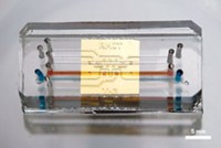
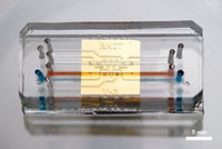
Chiu and his team developed an automated method that counts cancer cells quickly without damaging them. They selectively label the cancer cells with fluorescent dyes and then detect the cells as they travel through a microfluidic channel.
The only preparation the method requires is adding a set of three fluorescently labeled antibodies to a patient’s blood sample. Two antibodies recognize unique proteins found on the surfaces of breast cancer cells, EpCAM and cytokeratin. The scientists labeled the EpCAM antibody with a yellow fluorescent dye and the cytokeratin antibody with a red one. They also added a green dye to an antibody for CD45, a molecule absent from cancer cells. To reduce the chance of counting a cell that isn’t cancerous, Chiu’s method only counts a cell as cancerous if it glows yellow and red but not green.
The researchers tested the approach by mixing the fluorescent antibodies with blood samples from patients with breast cancer. They flowed the samples through a 3-cm-long microfluidic channel at a rate of more than 2 mL per hour. About halfway through the channel, two lasers shone on a 5-μm-long section of the channel, exciting any fluorescent dyes present. Using three small light detectors, the team monitored red, yellow, and green fluorescence and then counted cells as cancerous if the red and yellow but not the green detectors spotted a signal.
The researchers could analyze a single sample in a couple hours and detect as few as one cancer cell per milliliter of blood. Chiu’s team also compared their microfluidic method to the FDA-approved technique. They analyzed 90 blood samples from breast cancer patients with both methods. Their chip detected cancer cells in 82 of the samples, while the FDA-approved method only spotted the cells in about 40.
“This is definitely a big deal,” says Hang Lu of the Georgia Institute of Technology. She’s impressed by the method’s sensitivity and appreciates that the cells aren’t damaged in the process. Lu hopes that, in future studies, the researchers will capture the cancer cells to learn more about the differences between circulating tumor cells and cancer cells that stay put. Such information could help researchers better understand and possibly prevent metastasis, she says.


Join the conversation
Contact the reporter
Submit a Letter to the Editor for publication
Engage with us on Twitter