Advertisement
Grab your lab coat. Let's get started
Welcome!
Welcome!
Create an account below to get 6 C&EN articles per month, receive newsletters and more - all free.
It seems this is your first time logging in online. Please enter the following information to continue.
As an ACS member you automatically get access to this site. All we need is few more details to create your reading experience.
Not you? Sign in with a different account.
Not you? Sign in with a different account.
ERROR 1
ERROR 1
ERROR 2
ERROR 2
ERROR 2
ERROR 2
ERROR 2
Password and Confirm password must match.
If you have an ACS member number, please enter it here so we can link this account to your membership. (optional)
ERROR 2
ACS values your privacy. By submitting your information, you are gaining access to C&EN and subscribing to our weekly newsletter. We use the information you provide to make your reading experience better, and we will never sell your data to third party members.
Pharmaceuticals
Ferritin Taxis Anti-Cancer Agents To Tumors
Nanomedicine: Protein filled with light-sensitive molecules targets tumors for photodynamic therapy
by Leigh Krietsch Boerner
July 18, 2013

Cancer patients receiving photodynamic therapy have to deal with a unique side effect: They must avoid direct sunlight for one to two months. The reason is that light-sensitive compounds doctors inject into patients go to healthy as well as tumor tissue. Sunlight can activate these compounds, causing them to damage the healthy tissue. But now, scientists report that they can stuff large numbers of these molecules into the protein ferritin and deliver them right to tumor cells (ACS Nano 2013, DOI: 10.1021/nn402199g).
In photodynamic therapy (PDT), doctors inject porphyrin-based compounds into a patient, wait an appropriate amount of time for the molecules to reach the tumor, and then irradiate the area with a laser. These photosensitizers absorb the light, then transfer the energy to oxygen to create singlet oxygen, a reactive species that destroys cells nearby.
To make the porphyrins more water soluble and to better target them to tumors, chemists have tried enclosing the porphyrins inside nanoparticles, such as liposomes.
Nanoparticles tend to accumulate around tumors because the blood vessels that form around them are leaky. But those strategies have met with limited success because the researchers couldn’t load many light-sensitive molecules inside the particles, reducing the treatment’s effectiveness.
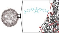
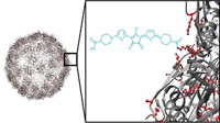
Jin Xie, a materials scientist at the University of Georgia, and his group wanted to find a soluble nanoparticle that could enclose large numbers of PDT molecules. So they encased the photosensitizer zinc hexadecafluorophthalocyanine in the protein ferritin, which they had explored previously as a carrier for chemotherapy drugs. Ferritin, found in almost every organism on earth, is made up of 24 subunits assembled into a hollow nanostructure. In cells, the cavity is filled with iron. But chemists can make hollow ferritin and easily fill it with other molecules. Even when filled, the protein nanostructure is just 18 nm in diameter and so is less likely to be cleared by the body’s immune system, Xie says.
To target the ferritin particles to tumors, the researchers added a small peptide called RGD4C to the outside of ferritin. This peptide binds to a receptor commonly found on tumor cells. To fill the particles, the team mixed the photosensitizer with the ferritin subunits. The particles could hold a large amount of the PDT molecules: The photosensitizers accounted for about 60% of the total particle mass. With past nanocarriers, the photosensitizers made up at most 10% of the total mass.
As one test of the ferritin particles, the team attached infrared dye molecules to the nanoparticles and injected them into mice that had human brain tumor cells placed under their skin. With an infrared camera, the researchers could see that the nanoparticles selectively accumulated at the tumor site and not in healthy tissue. After the researchers shined laser light on the tumor sites, tumor growth slowed significantly. The tumor growth rate in mice receiving filled ferritin particles and laser treatment was about 84% less than that of animals who did not receive the full treatment.
“The key advance is that they’re getting a really high payload capacity,” says Michael J. Sailor, a materials scientist at the University of California, San Diego. It’s this high loading that is responsible for the large amount of cell death, he adds. Sailor was also impressed by how well the targeting worked, which is not guaranteed, even with this known targeting peptide.


Join the conversation
Contact the reporter
Submit a Letter to the Editor for publication
Engage with us on Twitter