Advertisement
Grab your lab coat. Let's get started
Welcome!
Welcome!
Create an account below to get 6 C&EN articles per month, receive newsletters and more - all free.
It seems this is your first time logging in online. Please enter the following information to continue.
As an ACS member you automatically get access to this site. All we need is few more details to create your reading experience.
Not you? Sign in with a different account.
Not you? Sign in with a different account.
ERROR 1
ERROR 1
ERROR 2
ERROR 2
ERROR 2
ERROR 2
ERROR 2
Password and Confirm password must match.
If you have an ACS member number, please enter it here so we can link this account to your membership. (optional)
ERROR 2
ACS values your privacy. By submitting your information, you are gaining access to C&EN and subscribing to our weekly newsletter. We use the information you provide to make your reading experience better, and we will never sell your data to third party members.
Analytical Chemistry
Microfluidic Device Helps Scientists Capture Snapshots Of Biomolecule Interactions
Biophysics: By rapidly diluting solutions of biomolecule complexes, the device enables single-molecule studies before the molecules disassemble
by Naomi Lubick
July 2, 2013

Inside our cells, proteins and other biomolecules come together to form molecular complexes as part of the mechanisms that keep the cells running. These interactions sometimes are fleeting. For example, as new proteins are synthesized, the growing chain interacts off and on with other proteins called chaperones that ensure the new one folds correctly. A new microfluidic device could help researchers study short-lived molecular complexes involved in key biological processes, capturing snapshots of them one at a time (Anal. Chem. 2013, DOI: 10.1021/ac4010875).
Single-molecule fluorescence microscopy is a powerful tool for biochemists to study the short-lived behaviors and structure of biomolecules. In these experiments, scientists dilute solutions of their molecules of interest to very low concentrations so that they flow one at a time through the spotlight of a confocal microscope. The dilution takes time—usually a lot longer than some molecular complexes stay together. This timing issue makes the complexes difficult to study with single-molecule methods.
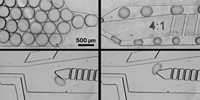
Researchers led by Tuomas P. J. Knowles and David Klenerman at the University of Cambridge designed the new microfluidic chip to solve this problem. The chip has two injection ports: one for a buffer and the other for a solution of the complex of interest. The liquids flow away from the ports through channels that twist back and forth like mountain switchbacks to prevent surges in pressure with each injection.
The solution of complexes gets diluted when it mixes with the stream of buffer at a series of four-way junctions. At each junction, the solution is diluted by one-tenth. In total, the chip passes the solution through five junctions in a few seconds, diluting the mixture 100,000-fold.
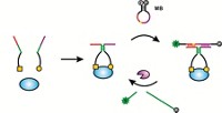
Mathew H. Horrocks, a graduate student in the team, tested the chip by diluting a solution of a pair of DNA strands labeled with fluorescent dyes. He added the device to a standard single-molecule fluorescence microscopy setup and looked for a fluorescent signal indicating that the strands remained together. The strands are so weakly associated that they normally fall apart in just 20 seconds. But with the chip in place, Horrocks could detect the characteristic fluorescent signal.

Because the dilutions happen quickly and require only one injection by the researcher, Klenerman thinks the circuitlike chip could lead to high-throughput, automated methods to analyze many samples. Sitting for long periods at a microscope, tending an experiment, can be tedious. “Our long term goal is to be able to run multiple single molecule experiments fully automatically without needing to actually be in the lab all day and all night,” he says.
The chip can be made with standard soft lithography techniques generally available in single-molecule research labs. With that in mind, the team provides blueprints for the chip on their website.
The team has developed a “clever, creative, and relatively simple” dilution method, says David A. Weitz, an applied physicist at Harvard University. While the Cambridge group will use the chip to look at proteins involved in Alzheimer’s and other neurodegenerative diseases, Weitz could see it being useful to study biomolecule interactions for pharmaceutical compounds. Plus, it’s easy to it into an experiment: Researchers could pluck the chip design off the web, fabricate it, and then plug the chip into any single-molecule confocal microscope setup.


Join the conversation
Contact the reporter
Submit a Letter to the Editor for publication
Engage with us on Twitter