Advertisement
Grab your lab coat. Let's get started
Welcome!
Welcome!
Create an account below to get 6 C&EN articles per month, receive newsletters and more - all free.
It seems this is your first time logging in online. Please enter the following information to continue.
As an ACS member you automatically get access to this site. All we need is few more details to create your reading experience.
Not you? Sign in with a different account.
Not you? Sign in with a different account.
ERROR 1
ERROR 1
ERROR 2
ERROR 2
ERROR 2
ERROR 2
ERROR 2
Password and Confirm password must match.
If you have an ACS member number, please enter it here so we can link this account to your membership. (optional)
ERROR 2
ACS values your privacy. By submitting your information, you are gaining access to C&EN and subscribing to our weekly newsletter. We use the information you provide to make your reading experience better, and we will never sell your data to third party members.
Analytical Chemistry
High-Throughput Method Analyzes Proteins Trapped In Preserved Tissues
Proteomics: A technique using antibody microarrays could find disease biomarkers in libraries of patient tissue samples
by Naomi Lubick
October 10, 2013

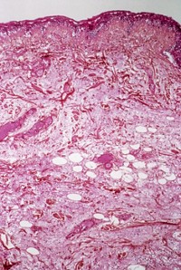
Hospitals and clinics routinely collect tissue samples from patient biopsies so that histologists can study them for visible signs of disease. These tissue samples are often preserved in formalin and fixed in paraffin, a preservation method that is optimal for analysis by techniques like microscopy but poorly suited for methods to characterize the proteins inside the tissues. Now, researchers have found a way to unlock the proteins from the paraffin matrix and identify them in a high-throughput manner (J. Proteome Res. 2013, DOI: 10.1021/pr4003245). The method may open up a treasure trove of data for researchers looking to find protein biomarkers of disease.
Millions of formalin-fixed, paraffin-embedded (FFPE) samples exist in tissue libraries all over the world. Because doctors know the diseases and outcomes for the patients corresponding to those samples, researchers think the libraries could be a great resource to connect protein biomarkers to certain diseases. Unfortunately, most methods to extract proteins from the samples fragment or unfold the proteins, making it difficult for typical protein assays to recognize the molecules. Several years ago, researchers at the National Cancer Institute (NCI) partnered with Expression Pathology Inc. (now OncoPlex Diagnostics) to get around this problem by analyzing the fragmented and unfolded proteins with mass spectrometry (Mol. Cell. Proteomics 2005, DOI: 10.1074/mcp.M500102-MCP200). But the method is slow, says Christer Wingren of Lund University, in Sweden. He and his team thought protein microarrays could lead to a method that was amenable to high-throughput screening for biomarkers.
Since 2000, Wingren’s group has been developing antibody microarray technology to identify and measure concentrations of biomarkers in clinical samples, like blood serum and plasma, using recombinant antibodies specific to certain proteins. With an improved extraction method that pulls out more whole proteins, Wingren thought the microarrays could identify those plus more of the protein fragments and unfolded proteins from the preserved tissue samples—and do it more quickly.
To analyze FFPE samples with antibody microarrays, Wingren and his colleagues first extracted whole proteins and protein fragments from samples collected at nearby Skåne University Hospital from healthy patients and those with breast cancer or one of two types of lymphoma. The researchers used xylene to remove the paraffin and then heated the samples to reverse the crosslinking of the proteins caused by the formalin. Then, after extracting the proteins with a commercial kit, the researchers added fluorescent labels to the proteins for later detection.
The team then applied the tissue extracts to antibody microarrays they fabricated using standard techniques. They used 35 antibodies to look for 11 proteins and seven protein fragments they knew existed in the tissues. When the researchers added the protein samples to the droplets, the antibodies bound to their targets, fixing the fluorescently labeled proteins onto the array. To determine the concentration of the proteins, the researchers measured the fluorescence intensities using a confocal microarray scanner and compared those measurements to samples that had been doped with known amounts of a specific target protein. The researchers could detect 16 of the 18 proteins and protein fragments, with a limit of detection of 1.25 to 5 μg/mL of total protein for 21 of the antibodies and 10 μg/mL for five others.
By pairing protein extraction methods with antibody microarrays, the new method can be used to detect proteins that may be difficult to analyze with mass spectrometry, says Timothy D. Veenstra, senior vice president of C2N Diagnostics in St. Louis, Mo., and one of the developers of the NCI’s FFPE extraction technique. However, one limitation of the technique is that it requires researchers to know which proteins they’re looking for so they can chose the right antibodies, says Antonio Lucacchini of the University of Pisa, in Italy. Still, the method’s increased sensitivity and amenability to rapid screens will help researchers turn the archives of FFPE samples around the world into data gold mines.


Join the conversation
Contact the reporter
Submit a Letter to the Editor for publication
Engage with us on Twitter