Advertisement
Grab your lab coat. Let's get started
Welcome!
Welcome!
Create an account below to get 6 C&EN articles per month, receive newsletters and more - all free.
It seems this is your first time logging in online. Please enter the following information to continue.
As an ACS member you automatically get access to this site. All we need is few more details to create your reading experience.
Not you? Sign in with a different account.
Not you? Sign in with a different account.
ERROR 1
ERROR 1
ERROR 2
ERROR 2
ERROR 2
ERROR 2
ERROR 2
Password and Confirm password must match.
If you have an ACS member number, please enter it here so we can link this account to your membership. (optional)
ERROR 2
ACS values your privacy. By submitting your information, you are gaining access to C&EN and subscribing to our weekly newsletter. We use the information you provide to make your reading experience better, and we will never sell your data to third party members.
Analytical Chemistry
Electrostatic Forces Make Ions Fly For Mass Spectrometry Imaging
Analytical Chemistry: Researchers develop mass spectrometry imaging method that requires little sample preparation
by Erika Gebel Berg
February 12, 2014

With mass spectrometry imaging, chemists can create maps of the locations of biological molecules within tissues and cells. Unfortunately, the dominant imaging methods that exist today often require extensive sample preparations, which limit the use and scope of the techniques. Researchers report a new mass spectrometry imaging technique based on electrostatic spray ionization that requires little manipulation of the sample before analysis (Anal. Chem. 2014, DOI: 10.1021/ac4031779).
Matrix-assisted laser desorption/ionization (MALDI) mass spectrometry is a current imaging favorite, thanks to the high-resolution images it can generate. However, the method has its drawbacks, such as the need to run the experiment inside a vacuum and to add a matrix material to the sample to help with ionization, says Hubert H. Girault of the Swiss Federal Institute of Technology (ETH), Lausanne. Finding a good matrix can be tricky, and running the experiments in a vacuum increases the complexity and the expense of the technique. Less complicated methods exist, such as desorption electrospray ionization (DESI), but they can’t compete with MALDI’s spatial resolution. Girault wanted to develop an imaging method that worked under ambient conditions, required little fuss to prepare samples, and generated high-resolution images.
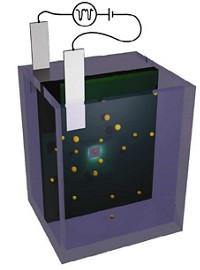
He and his colleagues recently developed a new ionization method called electrostatic spray ionization, or ESTASI (Anal. Chem. 2012, DOI: 10.1021/ac301332k). It differs from other forms of electrospray ionization techniques because it doesn’t require a probe to make direct contact with the sample’s surface. To create an imaging version of ESTASI, the researchers mount dry samples onto a moveable sample plate. The plate is sandwiched between a capillary attached to a mass spectrometer and a stainless steel electrode. The setup also includes a second capillary, 50 nm in diameter, that sits fixed in place above the sample. This wetting capillary delivers nanosized droplets of an acidic solution to the sample surface.
To generate images, researchers move the sample around underneath the wetting capillary. At each position, the electrode sitting below the plate generates high-voltage pulses, first at a positive voltage and then at a negative one, that ionizes molecules on the sample surface. The pulses also create a repulsive force that sends ions in droplets of the wetting solution flying away from the sample plate and into the capillary attached to the mass spectrometer. The positive pulse jettisons positive ions, while the negative pulse knocks off negative ions.
The researchers tested the mapping technique on melanoma cells, as well as patterns of spots of protein, peptides, and dye molecules. Under optimal conditions, the researchers could produce images with a resolution of 110 µm. That’s better than the typical resolution for DESI, which is between 180 and 200 µm, but not as good as MALDI’s resolution of about 10 µm. Girault hopes to improve the resolution by shrinking the wetting capillary, so it delivers smaller droplets, and by tweaking the composition of the solution it delivers.
Because the method requires no direct contact between the electrode and the sample, it could allow researchers to analyze samples in different formats, says Renato Zenobi of the Swiss Federal Institute of Technology (ETH), Zurich, who was not involved with the work. He is also impressed by the method’s easy and nondestructive sample preparation: “You could image a butterfly and then let it fly away after.”


Join the conversation
Contact the reporter
Submit a Letter to the Editor for publication
Engage with us on Twitter