Advertisement
Grab your lab coat. Let's get started
Welcome!
Welcome!
Create an account below to get 6 C&EN articles per month, receive newsletters and more - all free.
It seems this is your first time logging in online. Please enter the following information to continue.
As an ACS member you automatically get access to this site. All we need is few more details to create your reading experience.
Not you? Sign in with a different account.
Not you? Sign in with a different account.
ERROR 1
ERROR 1
ERROR 2
ERROR 2
ERROR 2
ERROR 2
ERROR 2
Password and Confirm password must match.
If you have an ACS member number, please enter it here so we can link this account to your membership. (optional)
ERROR 2
ACS values your privacy. By submitting your information, you are gaining access to C&EN and subscribing to our weekly newsletter. We use the information you provide to make your reading experience better, and we will never sell your data to third party members.
Biological Chemistry
First 3-D View Of The Body’s Pain-Sensing ‘Wasabi Receptor’
Biomolecular Structure: Snapshot of the TRPA1 ion channel, which senses chemical irritants, could lead to new pain and itch remedies
by Sarah Everts
April 13, 2015
| A version of this story appeared in
Volume 93, Issue 15

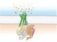
A wide variety of chemical irritants—including the molecules that give wasabi its kick and the toxic compounds that lend poison ivy its itch—activate the TRPA1 ion channel in the body. Switching on this channel leads to misery-producing pain, itch, or in the case of onions, tears. A structure of this important ion channel reported in Nature may pave the way to new analgesics that can tone down the protein when it signals too loudly (2015, DOI: 10.1038/nature14367).

“TRP” ion channels (pronounced “trip”) permit the passage of cations across cell membranes in plants, animals, and fungi. Humans have a whopping 27 different types of the channels. In 2013, David J. Julius and his colleagues at the University of California, San Francisco, used cryo-electron microscopy to solve the first structure of a member of this important family: TRPV1, which gets activated by capsaicin, the molecule that makes hot peppers hot. Julius’s team has now gotten a glimpse of TRPA1 using the same technique.
Although the researchers’ 4-Å-resolution structure isn’t detailed enough to pinpoint individual atoms, it does provide general features of the protein’s shape in its closed, or “off,” conformation, Julius says. The team found that four TRPA1 subunits assemble to form the ion channel and that this foursome requires an unusual phospholipid called inositol hexakisphosphate to be stable in cell membranes.
The TRPA1 structure also revealed that 80% of the protein’s mass is located outside the channel’s membrane-spanning core, on either side of the cell membrane. Some of this mass is involved in binding a wide assortment of chemical irritants. Curiously, rather than forming typical transient bonds, certain compounds that trigger TRPA1 activate the channel by forming covalent bonds to some of its cysteine or lysine amino acid residues, says David E. Clapham of Harvard Medical School in an associated commentary (Nature 2015, DOI: 10.1038/nature14383).
Opening and closing of the TRPA1 pore is likely regulated, at least in part, by a collection of so-called ankyrin repeats, amino acid sequences that hang from the channel down into the cell’s interior and form a docking platform for regulator proteins, Julius says. This docking platform has “a propeller-like structure, resembling the backs of four armadillos,” Clapham adds.
“In the future, more-detailed structures of TRPA1 in different conformations will reveal regulatory features, such as why the channel becomes sensitized and desensitized to calcium, and perhaps more importantly, how channel function can be blocked to treat asthma, inflammation, and pain,” Clapham notes.


Join the conversation
Contact the reporter
Submit a Letter to the Editor for publication
Engage with us on Twitter