Advertisement
Grab your lab coat. Let's get started
Welcome!
Welcome!
Create an account below to get 6 C&EN articles per month, receive newsletters and more - all free.
It seems this is your first time logging in online. Please enter the following information to continue.
As an ACS member you automatically get access to this site. All we need is few more details to create your reading experience.
Not you? Sign in with a different account.
Not you? Sign in with a different account.
ERROR 1
ERROR 1
ERROR 2
ERROR 2
ERROR 2
ERROR 2
ERROR 2
Password and Confirm password must match.
If you have an ACS member number, please enter it here so we can link this account to your membership. (optional)
ERROR 2
ACS values your privacy. By submitting your information, you are gaining access to C&EN and subscribing to our weekly newsletter. We use the information you provide to make your reading experience better, and we will never sell your data to third party members.
Analytical Chemistry
Raman Imaging In A Different Light
Microscopy: Modulating the frequency of laser beams allows researchers to pull out multiple Raman shifts to capture images of tissue samples
by Celia Henry Arnaud
November 2, 2015
| A version of this story appeared in
Volume 93, Issue 43
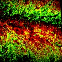
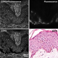
Ji-Xin Cheng and coworkers at Purdue University have developed an improved method for obtaining stimulated Raman scattering images of dense, highly scattering samples (Sci. Adv. 2015, DOI: 10.1126/sciadv.1500738). In the new method, the researchers modulate the pump laser beam at 16 frequencies in the megahertz range by dispersing the laser beam and scanning it onto a reflective pattern with 16 different densities. They then combine the pump beam with another laser beam, called the Stokes beam, and aim both at the sample. They record a time trace of the stimulated Raman signal from the sample and use a Fourier transform to demodulate the signal into 16 Raman shifts. With this approach, the Purdue team obtained stimulated Raman spectra of dimethyl sulfoxide that matched the spontaneous Raman spectra, even when pieces of dense tissue were placed in front of the detector to block the scattered photons from the sample. The team then mapped the active diffusion of α-tocopherol, also known as vitamin E, through mouse skin tissue. They also imaged intact human breast cancer tissue and live Caenorhabditis elegans worms.


Join the conversation
Contact the reporter
Submit a Letter to the Editor for publication
Engage with us on Twitter