Advertisement
Grab your lab coat. Let's get started
Welcome!
Welcome!
Create an account below to get 6 C&EN articles per month, receive newsletters and more - all free.
It seems this is your first time logging in online. Please enter the following information to continue.
As an ACS member you automatically get access to this site. All we need is few more details to create your reading experience.
Not you? Sign in with a different account.
Not you? Sign in with a different account.
ERROR 1
ERROR 1
ERROR 2
ERROR 2
ERROR 2
ERROR 2
ERROR 2
Password and Confirm password must match.
If you have an ACS member number, please enter it here so we can link this account to your membership. (optional)
ERROR 2
ACS values your privacy. By submitting your information, you are gaining access to C&EN and subscribing to our weekly newsletter. We use the information you provide to make your reading experience better, and we will never sell your data to third party members.
Synthesis
Structures of ‘hot’ cancer target solved
Knowing HDAC6’s configuration could help researchers design therapeutics
by Celia Henry Arnaud
July 27, 2016
| A version of this story appeared in
Volume 94, Issue 31

The enzyme HDAC6 is a “sizzling hot target for cancer chemotherapy,” says David W. Christianson, a chemist at the University of Pennsylvania who studies the enzyme. More formally known as histone deacetylase 6, this enzyme is responsible for removing acetyl groups from the protein tubulin, a structural component of a cell’s inner scaffold. Interfering with this deacetylation process can disrupt a cell’s ability to divide and transport nutrients, leading ultimately to cell death.

Because researchers would like to disrupt tumor cells this way, they’ve been looking for HDAC6 inhibitors as potential cancer treatments. So far, they have needed to search in the dark without a crystal structure to guide them—but not anymore.
Now two independent groups have solved structures of HDAC6 from zebrafish. Christianson and graduate student Yang Hai solved one set of crystal structures (Nat. Chem. Biol. 2016, DOI: 10.1038/nchembio.2134).They also solved one structure of human HDAC6, which showed that the zebrafish version is a suitable surrogate. The other set was solved by Patrick Matthias of the Friedrich Miescher Institute for Biomedical Research and coworkers (Nat. Chem. Biol. 2016, DOI: 10.1038/nchembio.2140).
Unlike other histone deacetylases, HDAC6 has two domains that catalyze acetyl group removal. Catalytic domain 2 (CD2) is the main site for deacetylation. Catalytic domain 1 (CD1), in contrast, is often called the “low activity” or “no activity” domain.
Christianson and Hai learned that, contrary to its nickname, CD1 can be quite active with the right substrate. CD1 has a lysine residue in its binding site where CD2 has a leucine. Using peptide substrates to model segments of tubulin, the researchers learned that CD1’s lysine serves as a “gatekeeper” that blocks the active site from all substrates except those that have an acetylated lysine at their carboxy terminus. The researchers aren’t yet sure why CD1 has that specificity in terms of its regulation of tubulin, Christianson says.
The team, however, was able to get snapshots of HDAC6’s catalytic mechanism. By replacing a histidine in the enzyme’s active site, the researchers were able to trap a tetrahedral intermediate of the deacetylation reaction. They also solved structures of HDAC6 with various substrates and inhibitors.
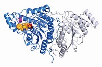
Both groups solved structures of the isolated catalytic domains. In addition, Matthias’s group solved a structure of CD1 and CD2 linked to one another. Together, the domains take on an ellipsoid shape with pseudo-twofold symmetry. “There is a large area of interaction between the two domains, with the linker on the outside,” Matthias says.
In addition, Matthias’s team found that HDAC6 gets its specificity for tubulin from a helix that is oriented differently than in other HDACs and from a flexible tryptophan in a loop connecting two of the enzyme’s helices.
“Both the Matthias and Christianson papers determine enzyme-inhibitor structures useful for pushing therapeutic discovery forward,” says Karen N. Allen, an enzyme researcher at Boston University. “These papers are chock-full of important information.”
This article has been translated into Spanish by Divulgame.org and can be found here.


Join the conversation
Contact the reporter
Submit a Letter to the Editor for publication
Engage with us on Twitter