Advertisement
Grab your lab coat. Let's get started
Welcome!
Welcome!
Create an account below to get 6 C&EN articles per month, receive newsletters and more - all free.
It seems this is your first time logging in online. Please enter the following information to continue.
As an ACS member you automatically get access to this site. All we need is few more details to create your reading experience.
Not you? Sign in with a different account.
Not you? Sign in with a different account.
ERROR 1
ERROR 1
ERROR 2
ERROR 2
ERROR 2
ERROR 2
ERROR 2
Password and Confirm password must match.
If you have an ACS member number, please enter it here so we can link this account to your membership. (optional)
ERROR 2
ACS values your privacy. By submitting your information, you are gaining access to C&EN and subscribing to our weekly newsletter. We use the information you provide to make your reading experience better, and we will never sell your data to third party members.
taxonomy
Getting a bigger picture with superresolution microscopy
Chip-based illumination enables simple microscopy set-up and wide field of view
by Celia Henry Arnaud
April 27, 2017
| A version of this story appeared in
Volume 95, Issue 18

Superresolution microscopy techniques allow researchers to observe objects tens of nanometers in size on or inside cells. But the methods can only keep an eye on patches 100 μm or less on a side at a time, making it difficult to image multiple cells simultaneously.
Now, by coupling a photonic chip with a standard optical microscope, researchers have achieved superresolution fluorescence microscopy with a simpler set-up and a wider field of view than conventional methods (Nat. Photonics 2017, DOI: 10.1038/nphoton.2017.55).
“You just need a basic microscope,” says Balpreet S. Ahluwalia of the Arctic University of Norway. “Our photonic chip technology can be retrofitted with any standard microscope to convert it into an optical nanoscope.” The chips allow Ahluwalia, Mark Schüttpelz of Bielefeld University, and coworkers to separate the illumination and detection pathways so the two don’t interfere with each other and enable the use of lenses with wider fields of view.
The waveguide chips direct laser light to the sample. Because the waveguides are made of high-refractive-index materials, the chips generate an intense evanescent field strong enough for the superresolution microscopy method called direct STORM (dSTORM).
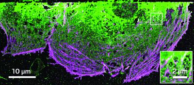
The researchers integrated the chip with a standard optical microscope equipped with either a 20X or a 60X objective lens. With the 60X objective, the spatial resolution was better than 50 nm. With the 20X objective, the spatial resolution was only 138 nm, but the field of view was an extraordinarily large 0.5 mm x 0.5 mm. They used the system to image structures in liver cells.
So far, the chips are limited to total internal reflectance fluorescence, or TIRF, excitation. “This limits the use to imaging structures up to 150–200 nm away from the waveguide surface,” Ahluwalia says. But that’s also a benefit, he says, because TIRF illumination allows researchers to look at thin slices with very little background.
Suliana Manley, a superresolution imaging expert at the Swiss Federal Institute of Technology, Lausanne, says the work “represents a significant advance in making TIRF microscopy more accessible, since it doesn’t require an expensive TIRF objective.” For superresolution imaging, membrane biologists are most likely to benefit from the technology, she says.
“Anyone interested in large-field-of-view imaging will benefit from this,” Ahluwalia says. “Using our photonic chip, we can generate with a slight reduction in spatial resolution a super-resolved image over approximately 100x larger area than what can be achieved today with commercial dSTORM microscopy.”
Advertisement
You might also like...


The power is now in your (nitrile gloved) hands
Sign up for a free account to get more articles. Or choose the ACS option that’s right for you.
Already have an ACS ID? Log in
Join the conversation
Contact the reporter
Submit a Letter to the Editor for publication
Engage with us on Twitter