Advertisement
Grab your lab coat. Let's get started
Welcome!
Welcome!
Create an account below to get 6 C&EN articles per month, receive newsletters and more - all free.
It seems this is your first time logging in online. Please enter the following information to continue.
As an ACS member you automatically get access to this site. All we need is few more details to create your reading experience.
Not you? Sign in with a different account.
Not you? Sign in with a different account.
ERROR 1
ERROR 1
ERROR 2
ERROR 2
ERROR 2
ERROR 2
ERROR 2
Password and Confirm password must match.
If you have an ACS member number, please enter it here so we can link this account to your membership. (optional)
ERROR 2
ACS values your privacy. By submitting your information, you are gaining access to C&EN and subscribing to our weekly newsletter. We use the information you provide to make your reading experience better, and we will never sell your data to third party members.
Biological Chemistry
Watching organelles bump into each other
Movies show how six types of the membrane-bound compartments interact simultaneously in live cells
by Stu Borman
May 24, 2017
| A version of this story appeared in
Volume 95, Issue 22

Cells have membrane-bound compartments, called organelles, that allow certain biochemical processes to proceed without interference from other cell chemistry. For example, lipids are synthesized in the endoplasmic reticulum, stored and transported as lipid droplets, oxidized in mitochondria and peroxisomes, and hydrolyzed and recycled in liposomes.
Various types of organelles move around in cells and bump into each other to transfer biomolecules and send and receive biochemical signals. But researchers have struggled to analyze the detailed ways in which organelles interact throughout cells over time.


Scientists have now made movies in whole cells showing simultaneous interactions of six types of organelles—lysosomes, mitochondria, endoplasmic reticulum, peroxisomes, Golgi, and lipid droplets (Nature 2017, DOI: 10.1038/nature22369). The work, by Jennifer Lippincott-Schwartz of Janelia Research Campus and coworkers, represents the most comprehensive analysis ever achieved of organelle motions and interactions, called the organelle interactome.
The researchers engineered monkey fibroblast cells to express four or five different-colored fluorescent proteins, each designed to localize in one type of organelle. For the other one or two organelles, the scientists added dyes to the cells known to target the desired compartments.
The team first used scanning confocal microscopy to monitor the fluorescent proteins and dyes point by point in thin slices of the cells. They analyzed organelle motions and contacts over 300 seconds and noted that the numbers of interactions decreased for most organelles in response to nocodazole, a drug that damages organelle-organizing microtubules. They also found that cell starvation increased lipid droplet interactions, likely to provide energy for the cell and restore reserves, and that excess fatty acids increased some lipid droplet interactions and decreased others to accommodate boosted fat metabolism.
Still, this point-by-point imaging with the scanning confocal microscope is very slow. To make three-dimensional videos of organelle interactions throughout whole cells, the researchers needed to speed up the imaging considerably. So they used lattice light-sheet microscopy to observe the fluorescent proteins and dyes simultaneously in cell slices. The scientists wrote software to distinguish each of the six monitored wavelengths and assembled those images to construct 3-D images from the slices. Lattice light-sheet microscopy, developed in 2015 by Janelia spectroscopist Eric Betzig, minimizes biological damage by illuminating thin slices of cells successively.
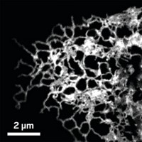
Among other findings, the movies show that the endoplasmic reticulum is the central organizer of the organelle interactome network and makes the most contacts with the other organelles.
The work is a breakthrough that “opens up wide-ranging opportunities for exploring the molecular mechanisms that underpin the organelle community’s dance,” writes Sang-Hee Shim of Korea University, who wrote a perspective accompanying the paper. “As the art of cell filmmaking matures, a new branch of systems biology based on images and video footage may emerge.”


Join the conversation
Contact the reporter
Submit a Letter to the Editor for publication
Engage with us on Twitter