Advertisement
Grab your lab coat. Let's get started
Welcome!
Welcome!
Create an account below to get 6 C&EN articles per month, receive newsletters and more - all free.
It seems this is your first time logging in online. Please enter the following information to continue.
As an ACS member you automatically get access to this site. All we need is few more details to create your reading experience.
Not you? Sign in with a different account.
Not you? Sign in with a different account.
ERROR 1
ERROR 1
ERROR 2
ERROR 2
ERROR 2
ERROR 2
ERROR 2
Password and Confirm password must match.
If you have an ACS member number, please enter it here so we can link this account to your membership. (optional)
ERROR 2
ACS values your privacy. By submitting your information, you are gaining access to C&EN and subscribing to our weekly newsletter. We use the information you provide to make your reading experience better, and we will never sell your data to third party members.
Biotechnology
Engineered cells detect tiny tumors in mice, paving the way for immunodiagnostics
Stanford researchers and a new biotech start-up called Earli are designing immune cells that release synthetic biomarkers for early cancer detection
by Ryan Cross
March 19, 2019
| A version of this story appeared in
Volume 97, Issue 12

Detecting cancer early is difficult. Imaging approaches, like positron emission tomography (PET), can’t spot tumors that are smaller than a few millimeters in diameter, and blood-based diagnostics struggle with parsing the minute quantities of biomarkers that signal cancer’s presence from the millions of other molecules in the blood. To make matters worse, cancer researchers struggle to find biomarkers that only cancer cells make.
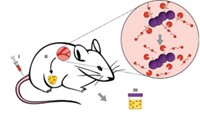
It’s a problem that Sanjiv S. Gambhir knows well. The cancer researcher and Radiology Department chair at Stanford University School of Medicine has spent the past five years “trying to flip the problem around.” His lab has engineered immune cells to produce glowing protein biomarkers when they bump into cancer cells. Studies in mice suggest this method could spot tiny tumors earlier than existing imaging and blood biomarker approaches (Nat. Biotechnol. 2019, DOI: 10.1038/s41587-019-0064-8).
Gambhir imagines that his cancer-sniffing cells could be combined with cell-based immunotherapies, which are engineered to kill cancer. “It sets the stage for an entirely new area of immunodiagnostics,” he says.
Although once the stuff of science fiction, engineered cells are on their way towards becoming a major pillar of the drug industry. The first two engineered cell therapies for cancer approved for sale in the US—Kymriah from Novartis and Yescarta from Gilead—catalyzed major investments in cell-therapy companies. The number of cancer cell therapies in development is surging, and the US Food and Drug Administration anticipates a tsunami of cell-therapy applications in the coming years.
Of course, these commercial cell therapies must first find cancer to kill it. But the current designs for detecting cancer are crude. Kymriah and Yescarta both attack any cell hoisting a protein marker called CD19 on their membranes. That strategy makes these cells great at killing certain kinds of leukemia and lymphoma cancers, but in the process, they also wipe out healthy white blood cells bearing CD19.
Gambhir’s cancer-sniffing cells take a different approach. While Kymriah and Yescarta are both made from a kind of immune cell called a T cell, Gambhir decided that a class of cells called macrophages were better for the job, since they naturally hone in on tumors in the body. “We don’t need to educate them to do that,” Gambhir says.
The Stanford team wanted to engineer these macrophages to secrete a bioluminescent protein called a luciferase when they found themselves inside a tumor. The use of such glowing proteins—which come from fireflies, microorganisms, and sea creatures—is common in research labs. The hard part was designing a genetic switch that would flip on to produce luciferase only when the macrophages come into contact with cancer.
To build that switch, Gambhir’s lab looked for genes in the macrophages that had high expression levels when the immune cells infiltrated tumors in mice, compared with when the cells just moved throughout the rodents’ bodies. They found that expression levels of one gene, called Arg1, jumped 200-fold in the tumor-honing macrophages. The team took part of the Arg1 gene called the promoter—the genetic code that turns the gene on—and stuck it in front of a gene from a small crustacean called Gaussia princeps that makes the glowing luciferase protein.
Advertisement
The team picked that copepod protein because cells both release some of it into their surroundings and hold onto some of it, Gambhir says. So doctors could look for luciferase in the blood or urine of patients to help diagnose cancer, and a follow-up PET scan could reveal the tumor’s location, thanks to the accumulation of luciferase. The team showed that the method could detect tumors as small as 25 to 50 mm3, or about the size of a pencil eraser.
Gambhir recalls that when he first submitted his study for publication at Nature Biotechnology over a year ago, the reviewers were “stunned that this could be made to work in a living animal.” But they weren’t convinced that it would outperform other approaches for early cancer detection. So Gambhir’s lab put their method to the test. They compared their engineered cells to an increasingly popular approach called cell-free DNA, which detects tumors by sequencing tiny bits of DNA that cancer cells release into the blood. The cell-free DNA approach couldn’t detect tumors until they were 1,500 to 2,000 mm3 in volume.
Victor E. Velculescu, a cancer biologist who studies cell-free DNA cancer diagnostics at the Johns Hopkins University School of Medicine, thinks Gambhir’s approach is promising. “It has advantages over existing methods in terms of detecting small lesions,” he says, but more studies will be needed to prove the approach works in humans.
Eleftherios Diamandis and Clare Fiala from the University of Toronto agree that the new approach is promising, but they also point out several caveats. Since the cell sensors were tested only in mice, “the conclusions about sensitivity or superiority over existing human cancer diagnostics are all speculation,” until tests are performed in humans, they say. Furthermore, people shouldn’t read too much into the small size of tumors detected because a 50 mm3 tumor in a mouse is equivalent to a tumor the size of two oranges in a human, they add.
Still, the idea of engineered cell sensors has attracted investor interest. Gambhir has licensed the technology to a new San Francisco–based start-up called Earli, which is backed by the tech-focused venture capital firm Andreessen Horowitz, which is also an investor in the cell-free DNA start-up called Freenome. Earli and Andreessen Horowitz both declined interview requests.
“This paper is an exciting beginning,” says Kole Roybal, a cell engineer at the University of California, San Francisco, who is designing cell sensors to spot what he calls precancerous states. But he notes that cell sensors alone may be impractical. “I think this is unlikely to be viable from a cost perspective unless the cells sense and treat the disease.”
Gambhir thinks that his cancer detecting approach could lead to therapies that kill cancer. “Right now, it seems a bit out there, but if cell therapies catch on, we can envision a time where you get an immunization with these cells,” he explains. The cells would live in you forever, detect cancer when it appears, and then kill it.
In the meantime, Gambhir’s lab is working on making the cell sensors as generalizable as possible, but he says he wouldn’t be surprised if some kinds of tumors can escape the engineered macrophages’ detection. “With any biological thing, it is absolutely hard to find 100% specificity,” he says.
In fact, his engineered cancer-spotting cells also emit luciferase when they find a wound in the body, because that environment also ramps up Arg1 activity. But Gambhir stresses that this experiment is a proof of principle. “It is certainly not done and ready for humans, it will require several years of work, but it sets up this nice strategy of hijacking immune cells,” Gambhir says. “I hope it leads to real tools that will help people someday.”


Join the conversation
Contact the reporter
Submit a Letter to the Editor for publication
Engage with us on Twitter