Advertisement
Grab your lab coat. Let's get started
Welcome!
Welcome!
Create an account below to get 6 C&EN articles per month, receive newsletters and more - all free.
It seems this is your first time logging in online. Please enter the following information to continue.
As an ACS member you automatically get access to this site. All we need is few more details to create your reading experience.
Not you? Sign in with a different account.
Not you? Sign in with a different account.
ERROR 1
ERROR 1
ERROR 2
ERROR 2
ERROR 2
ERROR 2
ERROR 2
Password and Confirm password must match.
If you have an ACS member number, please enter it here so we can link this account to your membership. (optional)
ERROR 2
ACS values your privacy. By submitting your information, you are gaining access to C&EN and subscribing to our weekly newsletter. We use the information you provide to make your reading experience better, and we will never sell your data to third party members.
Biotechnology
Ultrasound imaging for biochemistry
Noninvasive technique allows scientists to visualize biomolecular processes in tissues
by Laura Howes
July 16, 2020
| A version of this story appeared in
Volume 98, Issue 28

Fluorescent markers for imaging biomolecules have transformed science, but they have a major limitation. Light doesn’t penetrate well through tissue, so biomolecules deeper in the body have remained invisible. Now a team led by Mikhail Shapiro at the California Institute of Technology has developed a way to “hear” molecular processes: tunable acoustic biosensors that can be used to track biological processes pretty much anywhere within the body using ultrasound.
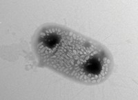
The biosensors are balloon-like nanoparticles that vibrate in response to ultrasound waves. These gas vesicles comprise a protein shell enclosing an air-filled compartment. Their widths are between 45 and 250 nm, and they are several hundred nanometers long. Shapiro’s team has engineered the tiny molecular balloons to detect protease activity. These enzymes chew up one of the proteins that makes the nanoparticles, changing their acoustic properties. That means researchers can use the nanoparticles to detect protease activity (Nature Chem. Biol. 2020, DOI: 10.1038/s41589-020-0591-0). The researchers showed the biosensors work inside bacteria and mice.
The key, Shapiro says, was realizing that the stiffness of the protein vesicles affected their acoustic properties. The more flexible the gas vesicles are, the more they scatter ultrasound. Introducing a protease-specific sequence into one of the vesicle components, a protein called GvpC, meant the signal changed if the protease enzyme was present. This change is analogous to how fluorescent tags show enzyme activity.
Lennart Verhagen, who investigates how ultrasound can modulate areas deep in the brain at Radboud University, says the new work is “beautiful.” Being able to image biological processes in deep tissue “has been long desired,” he says. “I expect it to be rapidly adopted by many other labs.”
Shapiro’s lab is now working to induce changes in the gas vesicles with other enzymes and biomolecules so they can also be detected via ultrasound imaging. With further development, he says, “the vision is that one day every biology lab will have an ultrasound setup next to the microscope.”
Correction
This story was updated on July 16, 2020, to correct the name of the protein in the nanoparticles. It is GvpC, not GdpC. Also, the figure's credit was absent. The figure was provided by Mikhail G. Shapiro.


Join the conversation
Contact the reporter
Submit a Letter to the Editor for publication
Engage with us on Twitter