Advertisement
Grab your lab coat. Let's get started
Welcome!
Welcome!
Create an account below to get 6 C&EN articles per month, receive newsletters and more - all free.
It seems this is your first time logging in online. Please enter the following information to continue.
As an ACS member you automatically get access to this site. All we need is few more details to create your reading experience.
Not you? Sign in with a different account.
Not you? Sign in with a different account.
ERROR 1
ERROR 1
ERROR 2
ERROR 2
ERROR 2
ERROR 2
ERROR 2
Password and Confirm password must match.
If you have an ACS member number, please enter it here so we can link this account to your membership. (optional)
ERROR 2
ACS values your privacy. By submitting your information, you are gaining access to C&EN and subscribing to our weekly newsletter. We use the information you provide to make your reading experience better, and we will never sell your data to third party members.
Cancer
Chemistry In Pictures
Chemistry in Pictures: Chip off the ol’ node
by Alexandra A. Taylor
October 19, 2020

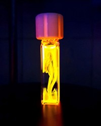
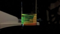
Microfluidic tissue-on-a-chip devices help researchers study drug toxicity and model the effects of flow conditions on cell behavior. While obtaining her PhD from the University of Illinois at Urbana-Champaign, Parinaz Fathi conducted research at the National Institute of Standards and Technology using this lymphatic vessel chip. Here the chip’s channels, which measure 1.75–5.5 mm wide, are filled with the fluorescent pigment biliverdin to make them stand out. Breast cancer tumors often metastasize to lymph nodes, and Fathi and coworkers used this chip to explore the effects of shear flow, or flow induced by a force, on cell alignment and cytokine production. “The health of the system, the flow conditions, and the existence of cytokines that encourage metastasis formation all interact with each other,” Fathi explains. She has since completed her PhD and now researches nanoparticle-based cancer immunotherapies as a postdoc at the National Institutes of Health.
Submitted by Parinaz Fathi (@ParinazFathi). Read the paper at ACS Applied Bio Materials 2020, DOI: 10.1021/acsabm.0c00609.
Do science. Take pictures. Win money. Enter our photo contest here.


Join the conversation
Contact the reporter
Submit a Letter to the Editor for publication
Engage with us on Twitter