Advertisement
Grab your lab coat. Let's get started
Welcome!
Welcome!
Create an account below to get 6 C&EN articles per month, receive newsletters and more - all free.
It seems this is your first time logging in online. Please enter the following information to continue.
As an ACS member you automatically get access to this site. All we need is few more details to create your reading experience.
Not you? Sign in with a different account.
Not you? Sign in with a different account.
ERROR 1
ERROR 1
ERROR 2
ERROR 2
ERROR 2
ERROR 2
ERROR 2
Password and Confirm password must match.
If you have an ACS member number, please enter it here so we can link this account to your membership. (optional)
ERROR 2
ACS values your privacy. By submitting your information, you are gaining access to C&EN and subscribing to our weekly newsletter. We use the information you provide to make your reading experience better, and we will never sell your data to third party members.
Microbiome
Lab chip models gut microbiome diversity
Intestine-on-a-chip device cultures both aerobic and anaerobic bacteria, could help researchers study how drugs interact with gut microbes
by Celia Henry Arnaud
May 15, 2019
| A version of this story appeared in
Volume 97, Issue 20
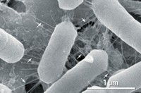
The human gut microbiome includes many types of bacteria that thrive in a variety of conditions. Some need oxygen—as do human cells—and others can’t tolerate high oxygen levels. Coming up with an experimental system that can mimic the human gut and meet the needs of both aerobic and anaerobic bacteria, as well as the host human cells, has been difficult. Such a device could help researchers study the interactions between gut microbes and our intestines, as well as understand how drugs interact with these bacteria.
“The challenge we all face in trying to build models that study the interface of gut bacteria and eukaryotic cells is providing an environment where both can thrive,” says Robert A. Britton, a microbiologist at Baylor College of Medicine.
A team led by Donald E. Ingber of the Wyss Institute for Biologically Inspired Engineering at Harvard University, has now developed a device that does just that. Ingber and his colleague have developed a microfluidic intestine-on-a-chip that can sustain aerobic and anaerobic bacteria in the same device (Nat. Biomed. Eng. 2019, DOI: 10.1038/s41551-019-0397-0).
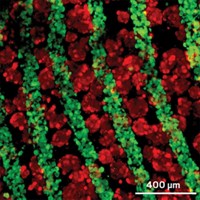
The 2-channel device has vascular endothelial cells in the lower channel. The upper channel has human intestinal epithelial cells that form fingerlike villi reminiscent of structures in the small intestine. Separating the two channels is a porous membrane coated with extracellular matrix, which mimics the biological scaffold that supports the two types of cells in our guts. Perfusing oxygenated cell-culture medium through the endothelium-lined channel while housing the device in a nitrogen chamber, allows the researchers to maintain the desired oxygen gradient, running from 20% to <0.5% oxygen, to keep all the cells happy.
The researchers added bacteria from human stool samples to the upper channel and cultured everything together. The device sustained a diverse population of more than 200 types of microbes with an abundance of the bacteria reflecting what’s typically observed in human stool samples.
“Most of what we know about the microbiome in medicine is due to genomic and metagenomic analysis, which is really guilt-by-association and correlation,” Ingber says. The device is “a tool to get at what people are recognizing as being a previously unexplored part of medicine that can lead to new therapeutics and diagnostics,” he says.
But the device could be difficult for labs without microfluidics expertise to use, Baylor’s Britton says. Nonetheless, the work “provides an important proof of concept for long-term cultivation of microbes and epithelial cells that will help inform the development of other, more accessible, model systems,” Britton says.


Join the conversation
Contact the reporter
Submit a Letter to the Editor for publication
Engage with us on Twitter