Advertisement
Grab your lab coat. Let's get started
Welcome!
Welcome!
Create an account below to get 6 C&EN articles per month, receive newsletters and more - all free.
It seems this is your first time logging in online. Please enter the following information to continue.
As an ACS member you automatically get access to this site. All we need is few more details to create your reading experience.
Not you? Sign in with a different account.
Not you? Sign in with a different account.
ERROR 1
ERROR 1
ERROR 2
ERROR 2
ERROR 2
ERROR 2
ERROR 2
Password and Confirm password must match.
If you have an ACS member number, please enter it here so we can link this account to your membership. (optional)
ERROR 2
ACS values your privacy. By submitting your information, you are gaining access to C&EN and subscribing to our weekly newsletter. We use the information you provide to make your reading experience better, and we will never sell your data to third party members.
Analytical Chemistry
Finding Crystallization Sweet Spots
Automated device mixes nanoliter quantities of membrane-protein components
by Mitch Jacoby
June 1, 2009
| A version of this story appeared in
Volume 87, Issue 22
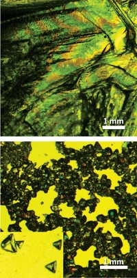
Membrane proteins play critical roles in cell signaling and energy transduction, so knowing their crystal structures could help scientists better understand how cells work while also pointing toward new treatments for diseases that involve these proteins. Relatively few membrane-protein structures have been determined, however, because they are notoriously difficult to crystallize and the pure proteins tend to be available only in minute quantities. What's more, today's crystallization methods do not allow experimental conditions to be easily varied, a feature that would hasten the identification of practical crystallization conditions for a particular protein. Now, Sarah L. Perry, Paul J. A. Kenis, and coworkers at the University of Illinois, Urbana-Champaign, have developed a device that enables them to screen crystallization conditions by mixing various compositions and concentrations of aqueous protein solutions and viscous lipids in 20-nanoliter batches (Cryst. Growth Des., DOI: 10.1021/cg900289d). The pneumatically actuated mixer uses just one-thousandth of the material consumed by today's microscale screening tools. In a proof-of-concept experiment, the team used the device to grow crystals of bacteriorhodopsin, a membrane protein commonly used for benchmarking.


Join the conversation
Contact the reporter
Submit a Letter to the Editor for publication
Engage with us on Twitter