Advertisement
Grab your lab coat. Let's get started
Welcome!
Welcome!
Create an account below to get 6 C&EN articles per month, receive newsletters and more - all free.
It seems this is your first time logging in online. Please enter the following information to continue.
As an ACS member you automatically get access to this site. All we need is few more details to create your reading experience.
Not you? Sign in with a different account.
Not you? Sign in with a different account.
ERROR 1
ERROR 1
ERROR 2
ERROR 2
ERROR 2
ERROR 2
ERROR 2
Password and Confirm password must match.
If you have an ACS member number, please enter it here so we can link this account to your membership. (optional)
ERROR 2
ACS values your privacy. By submitting your information, you are gaining access to C&EN and subscribing to our weekly newsletter. We use the information you provide to make your reading experience better, and we will never sell your data to third party members.
Biological Chemistry
Ebola's Clever Cloak
Structural Biology: Protein that hides viral RNA prevents immune system's detection of deadly virus
by Carmen Drahl
January 25, 2010
| A version of this story appeared in
Volume 88, Issue 4

A new molecular view sheds light on how the Ebola virus evades recognition by the immune system: It uses a cloaking mechanism to mask a telltale sign of its invasion (Nat. Struc. Mol. Biol., DOI: 10.1038/nsmb.1765). The finding could lead to treatments for Ebola hemorrhagic fever, the severe and often fatal disease that follows infection with the virus.
Ebola is a wily infectious agent for which there is no cure, explains Gaya K. Amarasinghe, a biochemist at Iowa State University. It leaves a calling card in cells upon infection: its double-stranded RNA. Usually, that RNA would trigger the human immune system. But the virus makes a protein called VP35 that masks the RNA, thereby squelching the immune response.
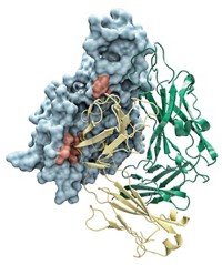
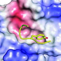
To learn more about how the masking works, Amarasinghe, virologist Christopher F. Basler of Mount Sinai School of Medicine, and colleagues determined the X-ray crystal structure of a double-stranded Ebola RNA bound to VP35 from the deadly Zaire species of the virus. They find that multiple copies of VP35 assemble to cloak the RNA. One copy uses a pocket of hydrophobic amino acids to recognize the RNA backbone and cap the end of the double-stranded RNA. Another copy binds to the backbone through a patch of basic amino acids. Changes to two of those basic residues are enough to destroy the virus’s disease-causing ability in guinea pigs, the team recently found (J. Virol., DOI: 10.1128/JVI.02459-09).
Last month, a team led by structural biologist Erica Ollmann Saphire of Scripps Research Institute detected the same unusual VP35-RNA binding in a less pathogenic species of Ebola (Proc. Natl. Acad. Sci. USA, DOI: 10.1073/pnas.0910547107). It’s gratifying that structures from different species of the virus, obtained under different conditions, show the same unorthodox mechanism, Ollmann Saphire says. “Of course, if you look at an unusual virus, you’re bound to find unusual things,” she adds.
Besides learning how the virus works, the team hopes the structural insights will fuel small-molecule drug discovery efforts for Ebola, which are currently lagging behind vaccine development, Basler says.


Join the conversation
Contact the reporter
Submit a Letter to the Editor for publication
Engage with us on Twitter