Advertisement
Grab your lab coat. Let's get started
Welcome!
Welcome!
Create an account below to get 6 C&EN articles per month, receive newsletters and more - all free.
It seems this is your first time logging in online. Please enter the following information to continue.
As an ACS member you automatically get access to this site. All we need is few more details to create your reading experience.
Not you? Sign in with a different account.
Not you? Sign in with a different account.
ERROR 1
ERROR 1
ERROR 2
ERROR 2
ERROR 2
ERROR 2
ERROR 2
Password and Confirm password must match.
If you have an ACS member number, please enter it here so we can link this account to your membership. (optional)
ERROR 2
ACS values your privacy. By submitting your information, you are gaining access to C&EN and subscribing to our weekly newsletter. We use the information you provide to make your reading experience better, and we will never sell your data to third party members.
Analytical Chemistry
Microfluidic Devices Capture Tumor Cells
Tools that run fluids through tiny channels detect scarce circulating tumor cells, a tactic that could aid cancer patients
by Celia Henry Arnaud
April 15, 2013
| A version of this story appeared in
Volume 91, Issue 15

One way cancer spreads is by shedding cells from the primary tumor that establish secondary tumors in other sites. The cells travel through the bloodstream, and more tumor cells in the blood hint at a worse outcome for the patient. The challenge is finding these circulating tumor cells (CTCs) against a background of regular cells. A single milliliter of blood contains approximately 5 million white blood cells and 5 billion red blood cells. How many CTCs are hiding in that crowd? Sometimes only one.
COVER STORY
Tumor Cells Snagged On The Fly
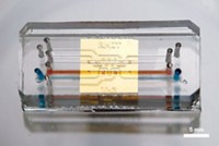
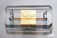
If doctors want to analyze these rare CTCs to assess the prognosis for cancer patients, they first have to fish for them. Microfluidic devices that pump liquids through tubes as thin as a hair to analyze them might be just the right kind of fishing rods. Speakers in two symposia at Pittcon focused on these devices and strategies for capturing the dangerous cells.
So far, just one method for counting CTCs has met with Food & Drug Administration approval, the CellSearch test sold by Veridex. In this test, cells are pulled out of blood samples with antibody-labeled magnetic beads and stained with targeted fluorescent dyes.
Scientists who are working toward new and improved tests to replace CellSearch start with a common definition: CTCs are nucleated cells that express two specific types of proteins on their surface, an epithelial protein cell adhesion molecule and one or more cytokeratins. And they don’t express the protein CD45, which is typically considered a marker for white blood cells.
As chemists and engineers are developing a variety of microfluidic CTC tools, they are observing that small changes in device geometry can make a big difference in capture efficiency. The researchers consistently find that their devices designed with new parameters in mind outperform the commercial Veridex system.
In one method, described by Daniel T. Chiu, a chemistry professor at the University of Washington, blood aliquots containing cells labeled with multiple fluorescently tagged antibodies are pumped through a microfluidic channel past a fluorescence microscope (Angew. Chem. Int. Ed., DOI: 10.1002/anie.201108695; Anal. Chem., DOI: 10.1021/ac400193b). Aliquots containing the rare cells are separated from the rest of the flow, and the rare cells are counted.

Another new method that combines microfluidics with magnetic capture to sort CTCs is the brainchild of Jean-Louis Viovy and coworkers at the Curie Institute, in Paris (Proc. Natl. Acad. Sci. USA, DOI: 10.1073/pnas.1001515107). These researchers pattern “magnetic anchors” on the bottom of a microfluidic channel and then flow antibody-labeled magnetic beads through the channel. In an applied magnetic field, the beads align with the anchors to form a hexagonal array of stacked beads. As a blood sample flows through the channel, the antibody-labeled beads pull the tumor cells out of the stream. Viovy and coworkers were able to capture not only circulating solid tumor cells but also lymphoma cells.
In another type of device, sticky “nano-Velcro” surfaces enable the detection, isolation, and analysis of CTCs. Hsian-Rong Tseng and coworkers at the University of California, Los Angeles, make the nano-Velcro from a nanostructured polymer fiber mesh, which snags nanostructures on cell surfaces. By coating the fibers with antibodies, the researchers can target specific cells. Tseng said his team captures more cells with the “hairy” surface than with a similarly functionalized smooth surface. The cells captured on the nano-Velcro chips can be treated and archived like conventional pathology samples, he said. His group used the chips to analyze individual circulating melanoma cells (Angew. Chem. Int. Ed., DOI: 10.1002/ange.201208452).

Some devices rely on geometric tweaks to improve CTC capture. For example, Steven A. Soper’s team at the University of North Carolina, Chapel Hill, makes devices with sinusoidal channels in the capture bed. The curved channels force cells close to the walls where they can be snagged by antibodies on the surface, he said (J. Am. Chem. Soc., DOI: 10.1021/ja8015022). His team has used the device to capture CTCs for analyses beyond simple counting, such as DNA analysis for known cancer-related mutations (Anal. Chem., DOI: 10.1021/ac103172y).
In yet another method, called geometrically enhanced differential immunocapture, antibody-coated posts are offset from one another in an array (PLoS ONE, DOI: 10.1371/journal.pone.0035976). The offset makes large cells like CTCs more likely to interact with the posts and be captured than they would be with an aligned array, said Steven M. Santana, a graduate student in Brian J. Kirby’s group at Cornell University.
Most of the new microfluidic devices rely on antibodies to capture or fluorescently label CTCs. But other options are available. Z. Hugh Fan, a professor at the University of Florida, uses ligand-binding nucleic acid sequences called aptamers, which he attaches to the channels in a microfluidic device (Anal. Chem., DOI: 10.1021/ac3005633). Fan isolates different types of CTCs by using aptamers with different specificities.
The new CTC-monitoring microfluidic technologies reported at Pittcon consistently outperform the Veridex system at detecting small numbers of cells cultured from cancer cell lines and spiked into whole blood. But Jeffrey J. Chalmers, a chemical engineering professor at Ohio State University, pointed out that cell lines are not the same as real CTCs in real patient samples.
In fact, Chalmers said, the Veridex definition of CTCs could exclude some relevant cells. Many CTCs undergo a transition in which they transform from epithelial cells into mesenchymal cells (a type of less-differentiated cells), which no longer have the characteristic markers. Chalmers is using microfluidic devices in an effort to expand the definition of CTCs to a broader range of cancer-associated cells with different surface markers.
The devices still need to win regulatory approval. But in the meantime, it’s clear they provide new ways to characterize CTCs. And armed with better information, researchers and their physician collaborators will be poised to make a difference for cancer patients, especially those who have cancers in which traditional imaging approaches don’t work well or who don’t have access to high-end technology.


Join the conversation
Contact the reporter
Submit a Letter to the Editor for publication
Engage with us on Twitter