Advertisement
Grab your lab coat. Let's get started
Welcome!
Welcome!
Create an account below to get 6 C&EN articles per month, receive newsletters and more - all free.
It seems this is your first time logging in online. Please enter the following information to continue.
As an ACS member you automatically get access to this site. All we need is few more details to create your reading experience.
Not you? Sign in with a different account.
Not you? Sign in with a different account.
ERROR 1
ERROR 1
ERROR 2
ERROR 2
ERROR 2
ERROR 2
ERROR 2
Password and Confirm password must match.
If you have an ACS member number, please enter it here so we can link this account to your membership. (optional)
ERROR 2
ACS values your privacy. By submitting your information, you are gaining access to C&EN and subscribing to our weekly newsletter. We use the information you provide to make your reading experience better, and we will never sell your data to third party members.
Analytical Chemistry
Mass spec imaging identifies tumor margins during brain surgery
Fast measurements distinguish among glioma, white matter, and gray matter, plus healthy versus diseased tissue
by Celia Henry Arnaud
June 19, 2017
| A version of this story appeared in
Volume 95, Issue 25
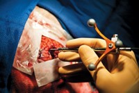
Glioma—a type of brain cancer that infiltrates surrounding tissue—is hard to distinguish visually and texturally from the surrounding healthy tissue. Surgeons need to remove as much of the cancer as possible while minimizing removal of healthy tissue. A team led by R. Graham Cooks of Purdue University and pathologist Eyas M. Hattab and neurosurgeon Aaron A. Cohen-Gadol of Indiana University School of Medicine have now used desorption electrospray ionization mass spectrometry in the operating room to assess tumor border during surgery to remove glioma (Proc. Natl. Acad. Sci. USA 2017, DOI: 10.1073/pnas.1706459114). The researchers obtained mass spectra of biopsied tissue smears in the operating room within three minutes, which is fast enough to be useful for making decisions during surgery. By using mass spec signals associated with membrane-derived lipids and N-acetylaspartate, the researchers were able to distinguish among glioma, white matter, and gray matter in the brain. In addition, they were able to detect a previously known glioma prognostic marker and determine the percentage of tumor cells in the biopsies. They followed the mass spectrometry determinations with conventional histopathological evaluations.


Join the conversation
Contact the reporter
Submit a Letter to the Editor for publication
Engage with us on Twitter