Advertisement
Grab your lab coat. Let's get started
Welcome!
Welcome!
Create an account below to get 6 C&EN articles per month, receive newsletters and more - all free.
It seems this is your first time logging in online. Please enter the following information to continue.
As an ACS member you automatically get access to this site. All we need is few more details to create your reading experience.
Not you? Sign in with a different account.
Not you? Sign in with a different account.
ERROR 1
ERROR 1
ERROR 2
ERROR 2
ERROR 2
ERROR 2
ERROR 2
Password and Confirm password must match.
If you have an ACS member number, please enter it here so we can link this account to your membership. (optional)
ERROR 2
ACS values your privacy. By submitting your information, you are gaining access to C&EN and subscribing to our weekly newsletter. We use the information you provide to make your reading experience better, and we will never sell your data to third party members.
Analytical Chemistry
Hydrogels Catch And Detect Cancer Cells
Bioanalytics: Researchers design hydrogels for use in a microfluidic device that indicate the presence of a protease overexpressed by cancer cells
by Leigh Krietsch Boerner
December 16, 2013

Cancer cells often leave a molecular calling card. They secrete an active version of the enzyme matrix metalloproteinase 9 (MM9) at higher levels than healthy cells do. With a new microfluidic technique, scientists can see if cancer cells are present by detecting MM9 (Anal. Chem. 2013, DOI: 10.1021/ac402660z). The method lets scientists detect as few as 11 cancer cells in a fluid sample.
MM9’s actions play a part in tumor cell invasion and metastasis. Because of the enzyme’s crucial role, researchers have developed other methods to measure protease concentration in cells but not in a real-time, high-throughput manner, says Dong-Sik Shin, a postdoctoral fellow in biomedical engineering at the University of California, Davis. In addition to speeding up cancer diagnostics, this would allow researchers to study how protease secretion by cancer cells changes in response to drugs.
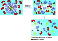
Shin, working with UC Davis professor Alexander Revzin and colleagues, designed a peptide that is cleaved by MM9. They attached a pair of molecules to each end of the peptide: one that fluoresces and another that quenches that fluorescence. The team placed the hydrogel in 100-µm-diameter circular wells in a microfluidic device, then attached the peptide to the gel with a thiol group. Cells from a liquid sample flow over the wells and get captured by an antibody in the hydrogel, Shin says.
When a cell producing MM9 lands in the well, the protease cleaves the peptide, and the quencher molecule floats away from the hydrogel, allowing the fluorescent molecule to glow. Higher levels of fluorescence mean greater amounts of protease, indicating the presence of cancer cells. The method is sensitive enough that the researchers can detect as few as 11 human lymphoma cells per well.


Join the conversation
Contact the reporter
Submit a Letter to the Editor for publication
Engage with us on Twitter