Advertisement
Grab your lab coat. Let's get started
Welcome!
Welcome!
Create an account below to get 6 C&EN articles per month, receive newsletters and more - all free.
It seems this is your first time logging in online. Please enter the following information to continue.
As an ACS member you automatically get access to this site. All we need is few more details to create your reading experience.
Not you? Sign in with a different account.
Not you? Sign in with a different account.
ERROR 1
ERROR 1
ERROR 2
ERROR 2
ERROR 2
ERROR 2
ERROR 2
Password and Confirm password must match.
If you have an ACS member number, please enter it here so we can link this account to your membership. (optional)
ERROR 2
ACS values your privacy. By submitting your information, you are gaining access to C&EN and subscribing to our weekly newsletter. We use the information you provide to make your reading experience better, and we will never sell your data to third party members.
Microscopy
Clear images and less damage from scanning transmission electron microscopes
New electronics tell the instrument when to block the electron beam, minimizing damaging effects
by Fionna Samuels
August 6, 2024

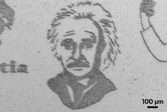
Scanning transmission electron microscopy, or STEM, is a powerful imaging technique that enables researchers to study a material’s morphology, composition, and bonding behavior at the angstrom scale. But the technique relies on a high-energy, focused beam of electrons, which ultimately damages the sample. Now a team led by researchers at Trinity College Dublin has developed a plug-and-play electronic system for STEM instruments that could change the way the devices generate images (Science 2024, DOI: 10.1126/science.ado8579).
The new system and approach were developed by physicists Lewys Jones and Jonathan Peters and their collaborators at Integrated Dynamic Electron Solutions (IDES) to connect a STEM instrument’s detectors to an electrostatic shutter placed in front of the instrument’s electron beam. As the beam scans a sample, it pauses point by point to collect data to generate each pixel of the final image. When a set number of electrons is detected scattering from each point, the shutter is triggered, and the electron beam is blocked, Jones says, protecting the sample from excessive damage. After a predetermined amount of time, the beam moves to the next point and is unblocked to collect another pixel’s worth of data.
“The electronics are constantly observing the signal, listening to and counting electron events and then making those logical decisions in real time,” Jones says.
The brightness of a pixel in the generated image reflects the time it took to detect the specified number of electron events from the sample at that location. Darker areas mean it took longer to detect the same number of electron events than for brighter areas. This approach is different from how STEM images have traditionally been generated—by bombarding each point with a continuous and damaging stream of electrons and translating the total number of electron events at each point into pixel brightness.
The new approach should reduce the destructive effects of electron beams, says Jingyue Liu of Arizona State University, a researcher who uses electron microscopes to study heterogeneous catalysts. Although this new technology will not completely eliminate the damage STEM does to a sample, Liu thinks it moves the field in the right direction. “If I had access to it, yeah, I’d like to test it on my sample,” he says.
The new technology is now available to researchers under the name Tempo, from turboTEM, a company Jones, Peters, and a colleague cofounded in 2022.
You might also like...

Advertisement
The power is now in your (nitrile gloved) hands
Sign up for a free account to get more articles. Or choose the ACS option that’s right for you.
Already have an ACS ID? Log in
Join the conversation
Contact the reporter
Submit a Letter to the Editor for publication
Engage with us on Twitter