Advertisement
Grab your lab coat. Let's get started
Welcome!
Welcome!
Create an account below to get 6 C&EN articles per month, receive newsletters and more - all free.
It seems this is your first time logging in online. Please enter the following information to continue.
As an ACS member you automatically get access to this site. All we need is few more details to create your reading experience.
Not you? Sign in with a different account.
Not you? Sign in with a different account.
ERROR 1
ERROR 1
ERROR 2
ERROR 2
ERROR 2
ERROR 2
ERROR 2
Password and Confirm password must match.
If you have an ACS member number, please enter it here so we can link this account to your membership. (optional)
ERROR 2
ACS values your privacy. By submitting your information, you are gaining access to C&EN and subscribing to our weekly newsletter. We use the information you provide to make your reading experience better, and we will never sell your data to third party members.
Analytical Chemistry
In Living Color
Nanoscale fluorescence imaging now in multicolor
by Celia Henry Arnaud
August 20, 2007
| A version of this story appeared in
Volume 85, Issue 34

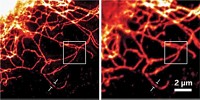
FLUORESCENCE IMAGING techniques sporting such catchy acronyms as STORM and PALM can zoom down to the nanometer scale (C&EN, Sept. 4, 2006, page 49), but capturing multicolor images with these methods remains a challenge because of the lack of suitable photoswitchable fluorescent probes. Now, two independent teams have demonstrated multicolor imaging with STORM and PALM, which both describe the same concept of super-resolution imaging.
"Imaging of two or more labels at the same time is critical for these methods to be useful in cell biology," says Stefan W. Hell, a biophysicist at Max Planck Institute for Biophysical Chemistry, in G??ttingen, Germany, and one of the team leaders. Multicolor imaging would allow researchers to look at many different cellular components at the same time. He and coworkers demonstrate two-color nanoscale microscopy with a variation of PALM and STORM (Appl. Phys. B 2007, 88, 161).
The other team, professor of chemistry and chemical biology Xiaowei Zhuang and coworkers at Harvard University and Howard Hughes Medical Institute, has identified a large family of photoswitchable probes based on cyanine dyes, which they use to generate multicolor STORM images (Science, DOI: 10.1126/science.1146598). Zhuang and coworkers rely on pairs of "activator" and "reporter" dyes that can be distinguished by either the reporter emission wavelength or the activation wavelength. By using several activators, the researchers selectively turn on subsets of reporters with different wavelengths of light. They have generated multicolor images of microtubules and clathrin-coated pits in cells at 20-30-nm resolution.
The number of available colors can be increased by combinatorially pairing the activators and reporters. "Even with just three reporters and three activators, I can potentially have nine colors," Zhuang says. "We expect that this will help not just super-resolution imaging but also normal imaging."
Hell's team uses fluorescent labels-a green photoswitchable fluorescent protein and a red dye-with well-separated excitation, emission, and switching characteristics. They likewise have collected multicolor images of the microtubule network.
STORM and PALM usually allow only a few reporter molecules to be turned on at the same time. Hell's group has now shown that a new rhodamine derivative allows images to be collected with a high density of tags (Angew. Chem. Int. Ed. 2007, 46, 6266). The rhodamine can also be turned on by two-photon absorption, which restricts the fluorescence to a single focal plane. By moving the focal plane, Hell and coworkers reconstruct three-dimensional images from optical slices.


Join the conversation
Contact the reporter
Submit a Letter to the Editor for publication
Engage with us on Twitter