Advertisement
Grab your lab coat. Let's get started
Welcome!
Welcome!
Create an account below to get 6 C&EN articles per month, receive newsletters and more - all free.
It seems this is your first time logging in online. Please enter the following information to continue.
As an ACS member you automatically get access to this site. All we need is few more details to create your reading experience.
Not you? Sign in with a different account.
Not you? Sign in with a different account.
ERROR 1
ERROR 1
ERROR 2
ERROR 2
ERROR 2
ERROR 2
ERROR 2
Password and Confirm password must match.
If you have an ACS member number, please enter it here so we can link this account to your membership. (optional)
ERROR 2
ACS values your privacy. By submitting your information, you are gaining access to C&EN and subscribing to our weekly newsletter. We use the information you provide to make your reading experience better, and we will never sell your data to third party members.
Pharmaceuticals
Specks Mark The Clot
Iron oxide nanoparticles functionalized with a fluorescent dye and a peptide light up newly formed clots for diagnostic imaging
by Aaron A. Rowe
June 8, 2009
| A version of this story appeared in
Volume 87, Issue 23
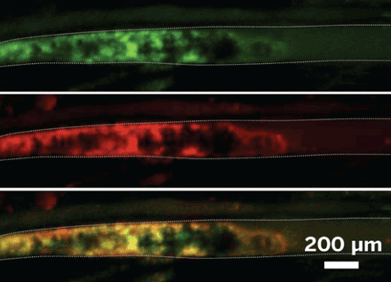
Iron oxide nanoparticles, when decorated with a fluorescent dye and the right peptide, can drift through blood vessels and light up newly formed clots to determine whether they are good candidates for treatment with thrombolytic drugs, according to a report by Jason R. McCarthy, Farouc A. Jaffer, and coworkers of Massachusetts General Hospital (Bioconjugate Chem., DOI: 10.1021/bc9001163). Removing those blockages with medication is risky because the same drug that clears one blood vessel may trigger serious bleeding in another area, such as the brain. To avoid those tragic side effects, the researchers proposed that functionalized nanoparticles could serve as magnetic resonance imaging contrast agents to help doctors decide whether a patient should receive clot-busting drugs. Bearing the dye molecules, the same nanoparticles make blockages shine for a catheter-based fluorescence microscope that can be used to watch a treated clot dissolve in real time. Key to the strategy, which the researchers tested in mice, is coupling the particles with a peptide that irreversibly binds factor XIIIa, a protein that is found only in fresh clots. The team plans further studies on the nanoparticles to evaluate thrombolytic drugs in vivo and to monitor clot formation on stents.


Join the conversation
Contact the reporter
Submit a Letter to the Editor for publication
Engage with us on Twitter