Advertisement
Grab your lab coat. Let's get started
Welcome!
Welcome!
Create an account below to get 6 C&EN articles per month, receive newsletters and more - all free.
It seems this is your first time logging in online. Please enter the following information to continue.
As an ACS member you automatically get access to this site. All we need is few more details to create your reading experience.
Not you? Sign in with a different account.
Not you? Sign in with a different account.
ERROR 1
ERROR 1
ERROR 2
ERROR 2
ERROR 2
ERROR 2
ERROR 2
Password and Confirm password must match.
If you have an ACS member number, please enter it here so we can link this account to your membership. (optional)
ERROR 2
ACS values your privacy. By submitting your information, you are gaining access to C&EN and subscribing to our weekly newsletter. We use the information you provide to make your reading experience better, and we will never sell your data to third party members.
Analytical Chemistry
Illuminating Tiny Bone Breaks
by Bethany Halford
November 30, 2009
| A version of this story appeared in
Volume 87, Issue 48
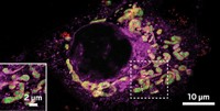
The first luminescent lanthanide contrast agent capable of imaging microcracks in bone has been developed by chemists in Ireland (J. Am. Chem. Soc., DOI: 10.1021/ja908006r). The cyclen-based europium compound could be used for bone structure analysis, lighting up barely visible fractures caused by repetitive loading and stress. Trinity College Dublin’s Thorfinnur Gunnlaugsson and coworkers constructed the contrast agent so that it contains three iminodiacetate moieties hanging off the cyclen ring. These groups selectively bind to exposed calcium sites in the damaged bone’s hydroxyapatite matrix, and a covalently linked naphthalene antenna sensitizes europium to its excited state. The researchers were able to differentiate polished and scratched bone four hours after exposure to the complex using confocal fluorescence laser-scanning microscopy. Gunnlaugsson tells C&EN that his group is currently developing the imaging agent for use in living systems.


Join the conversation
Contact the reporter
Submit a Letter to the Editor for publication
Engage with us on Twitter