Advertisement
Grab your lab coat. Let's get started
Welcome!
Welcome!
Create an account below to get 6 C&EN articles per month, receive newsletters and more - all free.
It seems this is your first time logging in online. Please enter the following information to continue.
As an ACS member you automatically get access to this site. All we need is few more details to create your reading experience.
Not you? Sign in with a different account.
Not you? Sign in with a different account.
ERROR 1
ERROR 1
ERROR 2
ERROR 2
ERROR 2
ERROR 2
ERROR 2
Password and Confirm password must match.
If you have an ACS member number, please enter it here so we can link this account to your membership. (optional)
ERROR 2
ACS values your privacy. By submitting your information, you are gaining access to C&EN and subscribing to our weekly newsletter. We use the information you provide to make your reading experience better, and we will never sell your data to third party members.
Pharmaceuticals
D-Protein Inhibits Key Drug Target
Drug Discovery: Mirror-image agent could lead to improved cancer and macular degeneration medicines
by Stu Borman
September 17, 2012
| A version of this story appeared in
Volume 90, Issue 38

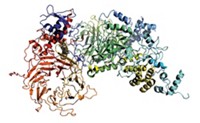
Researchers have used native chemical ligation, mirror-image phage display, and racemic protein crystallography to identify the first D-protein antagonist of vascular endothelial growth factor type A (VEGF-A).
The work could lead to improved cancer and macular degeneration drugs. Two injectable drugs, the anticancer drug Avastin and the macular degeneration agent Lucentis, are conventional L-protein VEGF-A antagonists. Because enzymes don’t recognize D-protein drugs, such drugs resist proteolytic breakdown and are potential oral medications. And the immune system doesn’t attack D-proteins because it doesn’t recognize them either.
The work was carried out by screening specialist Sachdev S. Sidhu of the University of Toronto, synthetic chemist Stephen B. H. Kent of the University of Chicago, and coworkers (Proc. Natl. Acad. Sci. USA, DOI: 10.1073/pnas.1210483109).
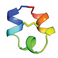
They used native chemical ligation to synthesize the 204-amino acid mirror image (D form) of VEGF-A and created phage libraries of variants of an L-protein called GB1 to identify one that recognized D-VEGF-A. They then synthesized the L-protein’s D form and found that it not only binds the natural L form of VEGF-A but also inhibits L-VEGF-A from binding its receptor—the desired mechanism of action of VEGF-A-targeted drugs. They used racemic protein crystallography to determine the structure of the ligand/VEGF-A complex.
Kent notes that the D-VEGF-A made in the study is twice the size of the largest D-protein previously synthesized and that the protein complex he and his coworkers analyzed structurally is more than 10 times the size of any determined previously by racemic crystallography.
Reflexion Pharmaceuticals, in San Francisco, is assessing the drug potential of the antagonist.
Several scientists told C&EN the study combines previous techniques but in a novel way. Stereochemistry expert Jay S. Siegel of the University of Zurich added that the researchers’ use of a symmetry argument to decide that the D form of the GB1-based protein would have features needed to bind and act as desired “is a really clever feature of the work.”


Join the conversation
Contact the reporter
Submit a Letter to the Editor for publication
Engage with us on Twitter