Advertisement
Grab your lab coat. Let's get started
Welcome!
Welcome!
Create an account below to get 6 C&EN articles per month, receive newsletters and more - all free.
It seems this is your first time logging in online. Please enter the following information to continue.
As an ACS member you automatically get access to this site. All we need is few more details to create your reading experience.
Not you? Sign in with a different account.
Not you? Sign in with a different account.
ERROR 1
ERROR 1
ERROR 2
ERROR 2
ERROR 2
ERROR 2
ERROR 2
Password and Confirm password must match.
If you have an ACS member number, please enter it here so we can link this account to your membership. (optional)
ERROR 2
ACS values your privacy. By submitting your information, you are gaining access to C&EN and subscribing to our weekly newsletter. We use the information you provide to make your reading experience better, and we will never sell your data to third party members.
Analytical Chemistry
Diagnosing Tuberculosis With A Nanotrap
Medical Diagnostics: Researchers couple nanopore technology with mass spectrometry to identify a peptide marker for infectious tuberculosis
by Erika Gebel Berg
February 3, 2014

The key to preventing the spread of tuberculosis—the second biggest killer among infectious diseases—is to rapidly identify people who are contagious and then keep them under quarantine. Yet current diagnostic methods are slow or unreliable. Now, by combining a nanoscale filter and mass spectrometry, researchers have developed a relatively speedy approach that could diagnose infectious cases of tuberculosis (Anal. Chem. 2014, DOI: 10.1021/ac4027669).
Many people around the world carry a dormant, latent form of Myobacterium tuberculosis, the tuberculosis bacterium, that isn’t contagious. So being able to differentiate it from contagious, active tuberculosis is critical, since people who carry the latent form needn’t be isolated. Quick screening approaches, such as the tuberculin skin test, can’t distinguish between the active and latent forms. The gold standard for diagnosing active tuberculosis requires taking a mucus sample from the lungs and growing the bacteria in a laboratory. The process “takes 10 days if you’re lucky, eight weeks if you’re not,” says Ye Hu of the Houston Methodist Research Institute. Hu and his team, who study disease-linked peptides, wanted to develop a faster screening method for the active form.
M. tuberculosis secretes a peptide called CFP-10, which is a “super-specific biomarker for diagnosing active tuberculosis,” Hu says. Mass spectrometry excels at identifying specific molecules, but detecting a small peptide like CFP-10 in complex biological samples can be like trying to find a needle in a haystack. That’s because signals from large proteins can obscure those from relatively small peptides, Hu says. In recent years, he has been developing nanopore thin films that act like elaborate filters to separate small peptides from large proteins for identification by mass spectrometry.
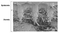
To make the nanopore thin films, the researchers combined silicate, an ethylene oxide and propylene oxide copolymer, and polypropylene glycol. They deposited the mixture on a 4-inch-square silicon wafer and allowed it to polymerize. Then, they heated the film to a balmy 450 °C for five hours, which removes the polypropylene glycol. What’s left looks a bit like an ant farm—a silica film with 5-nm-wide pores snaking throughout. The researchers covered the nanopore film with an adhesive plastic lid containing an array of 3-mm-diameter holes.
Next, the researchers grew M. tuberculosis cultures, filtered out the bacteria, and applied the remaining broth to the holes on top of the nanopore chip. Proteins too large to enter the nanopores remained on top of the film, while small peptides migrated into the porous film, Hu says. The researchers washed away the broth in the holes to get rid of the large proteins. They then applied the enzyme trypsin to the holes to cut up the peptides. Finally, the team washed the film with a buffer to pull out the chopped-up peptides.
The scientists performed mass spectrometry on the collected buffer and saw distinct peaks corresponding to CFP-10. The technique could detect peptide concentrations as low as 10 femtomolar, and the nanopore thin film isolated 90% of the CFP-10 in a sample. The whole process takes about nine hours, Hu says. He is currently testing whether his approach can isolate CFP-10 from patient serum samples.
Min Wu of the University of North Dakota says this overall approach offers hope for speeding up diagnosis of active tuberculosis. But he notes that mass spectrometry may not be available in parts of the world where tuberculosis detection is needed most.


Join the conversation
Contact the reporter
Submit a Letter to the Editor for publication
Engage with us on Twitter