Advertisement
Grab your lab coat. Let's get started
Welcome!
Welcome!
Create an account below to get 6 C&EN articles per month, receive newsletters and more - all free.
It seems this is your first time logging in online. Please enter the following information to continue.
As an ACS member you automatically get access to this site. All we need is few more details to create your reading experience.
Not you? Sign in with a different account.
Not you? Sign in with a different account.
ERROR 1
ERROR 1
ERROR 2
ERROR 2
ERROR 2
ERROR 2
ERROR 2
Password and Confirm password must match.
If you have an ACS member number, please enter it here so we can link this account to your membership. (optional)
ERROR 2
ACS values your privacy. By submitting your information, you are gaining access to C&EN and subscribing to our weekly newsletter. We use the information you provide to make your reading experience better, and we will never sell your data to third party members.
Analytical Chemistry
Protein Microarrays Go 3-D
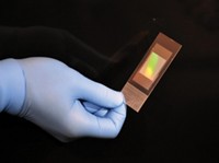
Biomarkers: A multilayered microarray detects multiple proteins in multiple microliter samples at once
by Erika Gebel Berg
April 25, 2014

With a protein microarray, researchers can find a specific protein in a biological sample, allowing them to diagnose a disease or unravel a physiological process. But sometimes one protein in one sample doesn’t provide enough information. To get the bigger picture, researchers have developed a simple and inexpensive three-dimensional protein microarray that can look for multiple proteins in multiple samples at the same time (Anal. Chem. 2014, DOI: 10.1021/ac501211m).
Currently, protein microarrays fall into one of two categories: forward phase and reverse phase. Forward-phase microarrays consist of a grid of spots coated with antibodies that each capture a certain protein in a sample. The method’s limitation is that it requires a large sample volume so that each spot can grab enough protein to produce a detectable signal, says Victoria de Lange, a graduate student working with János Vörös at the University of Zurich and Swiss Federal Institute of Technology, Zurich. With reverse-phase microarrays, researchers can work with small volumes. They dot different samples into an array and then probe the spots with a single antibody. Unfortunately, they can detect only one protein at a time.
“We felt something was missing in current protein microarray technology,” de Lange says. “We wanted to come up with a technology to combine the two techniques.” To make a microarray that can detect multiple proteins from low-volume samples, Vörös and de Lange designed a chip with 25 miniscule sample columns, consisting of multiple layers that each bind a different protein.
To build a prototype of the 3-D microarray, the researchers first printed five-by-five grids of 400-μm-diameter wax rings onto four sheets of nitrocellulose, a material that binds proteins tightly. The researchers then added antibodies to the rings in two of the layers: a mouse immunoglobulin G (mouse IgG) in one layer and a rabbit immunoglobulin G (rabbit IgG) in the other. They also added bovine serum albumin (BSA) to two other layers to serve as controls to determine if target proteins bound nonspecifically. The researchers then stacked and bound the layers on top of one another so that the wax rings all lined up. The wax rings created 25 sample columns that pass through each layer.
As a first test, the researchers prepared 1-μL samples containing the antigen for the mouse IgG, the antigen for the rabbit IgG, or a combination of the two, all tagged with a fluorescent dye. Each sample also had a high concentration of BSA to simulate serum. They pipetted the samples onto the top of the sample columns and centrifuged the microarrays, allowing the solutions to elute to the bottom. The researchers then took apart the microarray and looked to see which spots fluoresced on each layer. The mouse IgG layer glowed in only those spots where the researchers had added solutions containing its antigen. Similarly, the rabbit IgG layer produced signal only in the spots corresponding to the samples with the rabbit antigen. The BSA layers remained dark for all samples.
By testing samples with different concentrations of the proteins, the researchers determined that they could detect protein concentrations as low as 1.1 ng/mL. This range is clinically relevant, de Lange says, since cancer biomarkers are often in the ng/mL range. Also, she and Vörös point out that with a centrifuge and an ink printer to make the wax rings, any lab could build and use one of these 3-D microarrays.
“Wow, this is pretty nice,” says Julio A. Camarero of the University of Southern California. He says the device’s impressive feature is its ability to combine the advantages of both older protein microarray approaches. He wonders, though, how many different layers could be stacked together and thus how many proteins the approach could detect. De Lange says they tested up to 10 layers without a problem, and that she expects the 3-D microarrays could include up to 100 layers.


Join the conversation
Contact the reporter
Submit a Letter to the Editor for publication
Engage with us on Twitter