Advertisement
Grab your lab coat. Let's get started
Welcome!
Welcome!
Create an account below to get 6 C&EN articles per month, receive newsletters and more - all free.
It seems this is your first time logging in online. Please enter the following information to continue.
As an ACS member you automatically get access to this site. All we need is few more details to create your reading experience.
Not you? Sign in with a different account.
Not you? Sign in with a different account.
ERROR 1
ERROR 1
ERROR 2
ERROR 2
ERROR 2
ERROR 2
ERROR 2
Password and Confirm password must match.
If you have an ACS member number, please enter it here so we can link this account to your membership. (optional)
ERROR 2
ACS values your privacy. By submitting your information, you are gaining access to C&EN and subscribing to our weekly newsletter. We use the information you provide to make your reading experience better, and we will never sell your data to third party members.
Neuroscience
Chemistry In Pictures
Chemistry in Pictures: Brain in a dish
by Alexandra Taylor
November 21, 2018

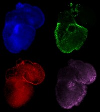
This micrograph shows a slice of a miniature, brainlike organoid. Organoids are three-dimensional cultures of cells grown in a lab to mimic an organ. Neuroscientist Alysson Muotri and his team at the University of California, San Diego, grew this one by stimulating human stem cells to develop into cortical tissue (neurons shown in red). They hope to one day use this method to examine how the brain develops and to model neurological conditions such as autism.
Credit: Muotri Lab
Do science. Take pictures. Win money. Enter our photo contest here.
Related C&EN Content:


Join the conversation
Contact the reporter
Submit a Letter to the Editor for publication
Engage with us on Twitter