Advertisement
Grab your lab coat. Let's get started
Welcome!
Welcome!
Create an account below to get 6 C&EN articles per month, receive newsletters and more - all free.
It seems this is your first time logging in online. Please enter the following information to continue.
As an ACS member you automatically get access to this site. All we need is few more details to create your reading experience.
Not you? Sign in with a different account.
Not you? Sign in with a different account.
ERROR 1
ERROR 1
ERROR 2
ERROR 2
ERROR 2
ERROR 2
ERROR 2
Password and Confirm password must match.
If you have an ACS member number, please enter it here so we can link this account to your membership. (optional)
ERROR 2
ACS values your privacy. By submitting your information, you are gaining access to C&EN and subscribing to our weekly newsletter. We use the information you provide to make your reading experience better, and we will never sell your data to third party members.
Business
Molecular Imaging
Companies set out to sharpen the in vivo perspective with new machines and novel contrast agents
by Vivien Marx, C&EN Northeast News Bureau
July 25, 2005
| A version of this story appeared in
Volume 83, Issue 30

The deep sea at around 3,000 meters is a dark, dark place. Many inhabitants of the ocean depths, such as the ferocious-looking barbeled dragonfish, light their way to food or a mate with a bioluminescent glow that cuts through the darkness around them.
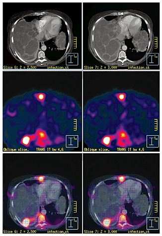
he body, too, is a dark place. Various imaging methods cut through that darkness noninvasively: fluorescence, ultrasound, computed tomography (CT), positron emission tomography
(PET), single-photon emission computed tomography (SPECT),and magnetic resonance imaging (MRI).
These optical, nuclear, and magnetic methods are now being empowered by new types of imaging agents. This synergy will make it possible to track the effectiveness of pharmaceuticals, treat disease, and monitor the response to therapies in novel ways. Companies in this area see interdisciplinary ventures as ways to both enhance drug development and change clinical medicine.
How well is a lead cardiovascular drug candidate working in vivo and over time? To what degree is a cancer drug causing tumor shrinkage soon after it is delivered? Frustratingly often, imaging results are too fuzzy for clear-cut answers. Biopsies are expensive and often not conclusive or practicable. The entire physiology of experimental animals cannot be efficiently monitored immediately after drug delivery and in the same animals over time to see if a drug is doing what it is designed for.
Imaging agents visually report from the scene of cellular events, such as when a drug latches onto its target. Or they can even deliver a therapeutic compound to a specific spot. Molecular imaging makes molecular processes visible, quantifiable, and trackable over time in a live animal or human, in "physiologically authentic environments," in the words of Sanjiv Sam Gambhir, who runs the molecular imaging program at Stanford University's medical school.
All imaging agents offer a readout about a particular region in the body. The long-used contrast agent barium sulfate, for example, uses physics as a stop sign. Blocked X-rays show up white on X-ray film, letting a physician visually examine the inner surface of the colon of a patient who has taken barium sulfate. Nuclear imaging, on the other hand, involves injecting a tracer--a diagnostic radiopharmaceutical--into the body and then detecting its radioactive emissions with a camera. Radioactive iodide, for example, is taken up in the thyroid and is helpful in examining thyroid disorders.

THE NEWER molecular imaging agents on the market and in development expand on such mechanisms and functionalities with chemistry. These agents, often called probes, aim to be smarter than their predecessors in that they amplify a specific cellular signal. Some agents only light up when an event of interest occurs.
According to Umar Mahmood of Massachusetts General Hospital's Center for Molecular Imaging Research (CMIR), disease is usually detected when structural changes in a tissue or organ have occurred. Molecular imaging may allow discovery of a "pre-disease state" in a patient when the first genomic and proteomic missteps in a pathway or cascade of events occur. Molecular imaging stands to show, in ways not previously possible, the dynamic underlying causes of disease while providing better treatment monitoring.
The technique might help "close the productivity gap" in drug development, as Janet Woodcock, acting deputy commissioner for operations at the Food & Drug Administration, has repeatedly pointed out in her speeches about the agency's Critical Path to New Medicinal Products initiative, intended to provide a dialogue between FDA and the drug industry.
Part of the pipeline problem, according to an FDA white paper called "Innovation or Stagnation," is that "the applied sciences needed for medical product development have not kept pace with the tremendous advances in the basic sciences." Imaging technologies can offer "powerful insights" into distribution, binding, and other facets of drugs or drug candidates, the report says. "New imaging technologies will ultimately contribute important biomarkers and surrogate endpoints, but how soon these new tools will be available for use will depend on the effort invested in developing them specifically for this purpose," the report states.
The application of imaging science is also big business. A study on the molecular imaging business published last month by market research firm Kalorama Information states that the movement of molecular imaging into the radiology laboratory "portends a big market opportunity" with large medical imaging companies recognizing their leverage in this field.
Companies such as Eli Lilly, GlaxoSmithKline, Novartis, and Bristol-Myers Squibb have set up medical imaging divisions and are integrating molecular imaging into their drug development efforts. In the preclinical stages, molecular imaging stands to become a high-content assay, reducing the number of experimental animals that need to be sacrificed because data is generated noninvasively. Imaging will help with the decision to take, or not take, a drug candidate into clinical trials and will offer another way to monitor efficacy once a drug is in trials.
Molecular imaging involves integrating new imaging technology with new imaging agents. This convergence is altering the focus of traditional imaging instrument makers. These firms are diving into "the wet stuff," as several imaging companies' executives call it: chemistry and biology.
"We are a combination of the traditional medical imaging device manufacturer [and] a diagnostic pharmaceutical company," says Alexander Tokman, general manager of global radiopharmacy and molecular imaging business at GE Healthcare. GE Healthcare, Philips Medical Systems, and Siemens Medical Solutions are, according to the Kalorama study, the top three market competitors in the imaging industry. GE says it has invested more than $160 million in the development of molecular imaging technology.
"The drug companies approach us to partner [and] to provide them with molecular imaging techniques, tools, and contrast agents to facilitate drug development," Tokman says. Philips and Siemens, too, are investing heavily in molecular imaging. "Molecular imaging serves as a great way of shortcutting the traditional pharma development model," says Ward Digby, director of molecular imaging at Siemens. Such imaging offers quicker feedback and "more insight into what is going on, first in animal models and later in human studies."
For the past 20 years, the big imaging technologies, or "modalities," have been mainly used on anatomy and not necessarily on a bodily process, Digby says. "The cool new growth area is telling something specific that is going on in the body that is related to disease," he says. "And you just can't do that very well with the anatomic agents." In his view, "the big emerging market is the pharma and biotech world realizing this is a great tool."
Some of the companies developing molecular imaging agents, such as Schering AG, Mallinckrodt, and Bracco, are also in the older contrast media field. Others, like the instrument makers, are newer arrivals, and others still are spin-offs from academic labs. All are eyeing the preclinical market in research and drug development as well as clinical applications.
According to Douglas A. Bakan, vice president of business development at Alerion Biomedical, a San Diego-based start-up that is developing magnetic resonance agents, the "easiest path to acceptance" for molecular imaging is in academic research. But, he says, one level up is big pharma, where decisions must be reached about taking a drug candidate through expensive trials or not. "The sooner we can give them that information and the more specific and accurate it can be, the better the chances that they can save time and money in the drug development process," Bakan says.
FOR INSTRUMENT MAKERS, the changed focus means embarking on unusual collaborative efforts to get an edge on licensing new technology and approaches. Given the rapid pace of change in biomedicine, the motivation for imaging companies to ally themselves in new ways is "part protective and part mercenary," says Raymond P. Thek, an attorney in the technology group of Lowenstein Sandler, a New Jersey-based law firm.
"The very way in which medical services are consumed is going to change," he says. Innovative activities are being pursued by a number of players, so the imaging instrument makers need to be a part of it, he says, or risk becoming "the Western Union in the age of the telephone."
For venture capitalists, the diagnostics field has historically lacked luster since it addresses markets that are smaller than those of therapeutics. For some venture capitalists, however, molecular imaging changes that perspective.
"What makes this particular field exciting is that you can very easily pair a therapeutic with a diagnostic," says Thomas C. Melzer, a managing director of RiverVest Venture Partners, a St. Louis-based venture-capital company. Because the cellular processes imaged by the probes are often the same processes that a therapeutic can target, a diagnostic imaging agent could be equipped to deliver a therapeutic compound directly to a diseased site. It is that combined power that makes this field "a very exciting business proposition," Melzer says.
In a talk at last year's American Association for Cancer Research Meeting, Markus Rudin, from the discovery technologies division at the Novartis Institutes for BioMedical Research in Basel, Switzerland, described molecular imaging as the "missing link" needed between animal imaging and human imaging. He is "very convinced" of the impact of molecular imaging on drug discovery and development. In a few years, he said, molecular imaging will become "a tool we cannot live without."
Using optical imaging, Rudin and colleagues Hans-Ulrich Gremlich, Martin Hintersteiner, and others just published their in vivo detection of amyloid-ß plaques (Nat. Biotechnol. 2005, 23, 577) These clumped bundles of peptides are a hallmark of Alzheimer's disease, for which cognitive tests are currently the main diagnostic.
The Novartis researchers synthesized and confirmed specific activity of a novel near-infrared fluorescence (NIRF) oxazine dye. In living mice, the dye crossed the blood-brain barrier, bound to the plaques in the brain, and gave off a fluorescence signal indicative of the plaque load. The Novartis group notes that this plaque-specific oxazine NIRF dye is "an attractive probe to noninvasively monitor disease progression in animal models of Alzheimer's disease and to evaluate the effectiveness of potential drugs."
As Ching H. Tung of Mass General's CMIR points out, optical imaging in live tissue is tricky because light is scattered and some of it absorbed by, for example, water or hemoglobin. NIRF in the 650- to 950-nm range offers a way to circumvent that drawback, Tung says. A number of near-infrared cyanine dyes are commercially available, with most of the fluorochromes showing similarity to a tribocyanine dye called indocyanine green.

THE NEXT GENERATION of optical imaging agents includes enzyme-activated optical imaging probes developed by CMIR scientists. The probes have a low fluorescence signal until they are activated by target enzymes such as proteases, after which their signal is amplified. Tung and his colleagues believe that such probes, which can be peptide- or polymer-based, show great promise for disease diagnosis and drug development.
Molecular imaging efforts to detect Alzheimer's disease and other neurological conditions are under way elsewhere; for example, in a partnership between GE Healthcare and Eli Lilly. Pittsburgh compound B (PIB), a thioflavin derivative developed at the University of Pittsburgh and licensed by GE, has been approved for use in Alzheimer's patients as a PET probe. According to Kalorama analyst Joseph Smith, a new Alzheimer's imaging agent such as PIB has "enormous market potential" and could reap yearly revenues of $1.8 billion.
Hardware--the physics of image capture--and software--the algorithms that construct an image from the captured data--cannot do the trick alone. As medical imaging companies move into molecular imaging, the idea is to be able to offer hardware as well as companion reagents. Instrument companies are starting to realize, according to Kalorama, "that the revenue potential of the agents is greater than the equipment sales themselves." Not only might these agents help sell more hardware, according to Smith, but a successful molecular imaging agent "could easily see $1 billion sales total during the lifetime of its patent."
The Novartis NIRF imaging project was completed with a Siemens fluorescence reflectance small-animal imager. That instrument was developed in collaboration with CMIR, says Siemens' Digby, where the development of new optical imaging agents is also being pursued for injection in small animals and "eventually, potentially in people," he says.
Advertisement
Siemens' partnership with CMIR, Digby explains, involves all the technologies of molecular imaging: optical imaging, magnetic resonance, and nuclear medicine. "It gives us a real head start if we are contributing to the development of the new tracer, developing the right software to evaluate it, and the hardware is tuned up right to the tracer," he says.
Digby joined Siemens a year ago from CTI Molecular Imaging, a longtime Siemens partner, and Siemens is just now completing the acquisition of CTI for $1 billion. CTI was devoted to PET products and had, for example, developed a modular automated radiosynthesis system with which chemists could synthesize a range of biomarkers such as fluorine-18 and carbon-11 radiolabeled compounds.
Siemens believes that preclinical imaging is increasingly important in assessing pharmacodynamics and pharmacokinetics. Many imaging instrument makers had been staying in their "established sandbox," Digby says, but interest in agents has grown immensely. He views Siemens' involvement in molecular imaging agents as "a real departure."
Digby sees an important present and an even more important future in PET. While PET may be a bit more complicated to use than SPECT and needs a cyclotron nearby to generate the necessary isotopes, he sees higher growth in PET than SPECT. "All molecular imaging techniques have to use relatively large molecular tags to generate the signal, except for PET," he says.
With SPECT, the most common isotope used is technetium, a large metal that must be chelated to the molecule of interest, Digby says. PET uses small isotopes--carbon, nitrogen, fluorine, oxygen--which permit the labeling of naturally occurring molecules--say, substituting fluorine for hydrogen--which is "a big intrinsic advantage," Digby explains.
The choice of agent and imaging modality depends, he adds, on the questions being asked. The challenge is that the specific targets are in the tissue at low concentrations, so agents must provide specificity and deliver a strong signal as well. Finding those agents requires investment.
Many industry observers credit GE as having given this corner of the instrumentation world a big life sciences push. GE's $9.5 billion acquisition in 2003 of Amersham, a diagnostics specialist, led to the formation of GE Healthcare. Other acquisitions followed, such as Enhanced Vision Systems and Suinsa. "We basically fine-tuned our vision to develop a combination of targeted diagnostics along with imaging equipment and information technologies," says Tokman of GE's new focus.
"GE's acquisition of Amersham has changed the whole playing field," says venture capitalist Melzer. "Philips and Siemens have to now think about how they want to respond to that."
Prior to the Amersham acquisition, the National Cancer Institute (NCI) had approached GE proposing to jointly develop a PET tracer. As Tokman explains, NCI has a pipeline of its own PET tracers because it is trying to expand the number of them on the market.
CURRENTLY, the most widely used PET radiopharmaceutical is the glucose analog 18F-FDG (fluoro-2-deoxy-D-glucose). Because tumors have an elevated glucose metabolism, 18F-FDG accumulates more in tumors than elsewhere. One idea is to develop a tracer that offers information complementary to glucose metabolism, namely cellular proliferation. That data can help better characterize tumors in vivo and improve the monitoring of cancer therapy response.
In the NCI collaboration, Tokman explains, GE scientists helped to develop the synthesis: producing the F-18 radioactive isotope in the firm's cyclotron and tagging it to thymidine to form the desired tracer, 18-fluorothymidine (FLT). Last November, GE and NCI filed a joint investigational new drug application for the compound with FDA, and it is now in clinical trials.
Ultimately, Tokman says, hospitals will all have molecular imaging centers that offer multidisciplinal expertise. Currently, a patient needs to wait months for a follow-up CT exam to determine if a therapy has been successful or needs to be changed. "FLT will be able to tell us if the therapy has been working after just a few days following treatment," he says.
Imaging companies stand to gain as molecular imaging makes inroads in drug discovery and in the clinic. Academic alliances are part of the scheme. At the Society for Nuclear Medicine's annual meeting last month, GE Healthcare announced a new five-year collaboration with the department of radiology at Stanford's medical school. The project brings together chemists and medical physicists, as well as clinicians and biologists, to create imaging technologies and molecular probes.
Stanford will house GE Healthcare instruments including a cyclotron, PET radiochemistry research equipment, and preclinical imaging technologies. Gambhir and his colleagues have approaches of interest for both optical and PET imaging agents, Tokman says. "We want to help him take that from development to the clinic."
Philips, too, has focused on academic collaborations in its molecular imaging endeavors and sees great promise in hybrid imaging technologies such as SPECT/CT and PET/CT.
In March, the company expanded a decadelong partnership with Washington University School of Medicine in St. Louis, notably in the laboratories of scientists Samuel A. Wickline and Gregory M. Lanza, who cofounded the spin-off firm Kereos to develop molecular imaging agents. Philips is supporting a center of applied nanomedicine at the medical school. It will belong to a biotechnology complex that is expected to be completed in 2006.
Philips has also joined up with Dow Chemical and Kereos in a Washington University project funded by NCI to develop and test novel molecular imaging agents for cancer as well as to develop the related nuclear imaging technology and image analysis techniques.
The researchers at Washington University are developing the target-specific nanoparticles, Philips is working out the imaging technology, and Dow scientists are figuring out how to attach radionuclides to the nanoparticles both for diagnostic and therapeutic purposes. Specifically, Dowpharma's ChelaMed radiopharmaceutical services unit has been working on new chelating molecules that can be used with Kereos' imaging agents. Kereos is responsible for evaluating the agents that result from this collaboration in order to prepare them for clinical trials, explains Robert A. Beardsley, the firm's chief executive officer.
According to Beardsley, approved molecular imaging agents are mainly nuclear--for example, a monoclonal antibody linked to a radionuclide. Their main disadvantage, he says, is a very high dose cost. "By the time you include the radiopharmacy and the monoclonal antibody and some other peculiarities of each agent, a dose may cost upwards of several thousand dollars."
While PET is a growing imaging technology, it faces some resolution challenges, in his view. The radioisotopes have a short half-life, and PET does not offer anatomical information unless coupled with a CT scan.
Kereos' imaging agents, which are geared toward MRI, are phospholipids with a perfluorocarbon core. The lipid tails associate against the surface of the perfluorocarbon, and the phosphate groups face out toward the water, Beardsley says. "We anchor into that phospholipid layer a targeting ligand to which we have conjugated a lipid tail," he says.
When the particle is injected, it can latch onto a specific site in the body, such as a tumor or atherosclerotic plaque, and be imaged at that precise location. Ten to 100 targeting ligands get the molecule to where it is supposed to go, and each particle can carry 100,000 or more payload molecules, he explains. The particles could potentially deliver therapeutics, whose effect could also be monitored noninvasively with imaging.

Kereos has "a two-way deal" with Bristol-Myers Squibb, Beardsley says. Kereos licensed an MRI agent targeted to fibrin for cardiovascular MRI, and the company has in-licensed a small-molecule ligand that targets a key hallmark of cancer, alpha v beta 3, an angiogenesis biomarker, and uses it as a targeting ligand. "We are able to take our particles to where there is a lot of alpha v beta 3 on the blood vessel wall," Beardsley says. This novel MRI agent in development is designed to be a biomarker for cancer that is also able to deliver a chemotherapeutic to the tumor site.
As F. David Rollo, chief medical officer at Philips Medical Systems, says, pretty much all of the molecular imaging agents are in development by academic spin-offs. Those firms will spend three to five years showing that their agent can localize to a specific spot in the body. "But when you go to the next level, trying to create the images for the clinical validation and the FDA approval, it turns out that there are some special requirements needed of the imaging device," he says.
Participants in the imaging field haven't done a good job of looking at how the imaging device and the agent work in combination, Rollo maintains. There are imaging agents that are approved but not widely used because the images obtained with them are not of acceptable quality. That challenge is where Philips has found an important niche, he says, collaborating with companies that either have an approved agent or one in late stages of development. "There's a real opportunity," he says, "to go back to the approved agents and show you can improve the image quality and therefore the usefulness of those agents."
Advertisement
The collaboration entails making the agent available to sites with Philips equipment. Philips collects the image data sets and works through the reconstruction algorithms to convert the data into an image, Rollo says. "By using different combinations of reconstruction algorithms and certain corrections such as attenuation and scatter corrections, we have found that we can, in fact, optimize the images," he says.

THAT R&D WORK, he says, reveals that when it comes to reconstruction algorithms, it is not one size fits all. Some molecular imaging agents are labeled with technetium-99m, others with iodine-123, and others still with indium-111, each with their own energies and scatter correction and attenuation correction requirements. Strategically, Philips has sought out companies with agents labeled with the different radioisotopes to work out ways to optimize imaging in each case.
ProstaScint, a monoclonal antibody-based prostate cancer imaging agent labeled with indium-111, was approved in 1997 but has not been used to the extent that its marketer, Cytogen, had expected, Rollo says. "We felt there was great potential for the agent if we could improve the image quality and better define the location of the areas of abnormality," he says.
With new algorithms, he and his colleagues were able to remove fuzziness in the appearance of the images, leading to a clearer outline of the lesion--the recurrent prostate cancer. That enhancement offers "the opportunity to change the method of therapy," Rollo says. Although ProstaScint isn't a blockbuster, its sales have grown steadily, rising from $4.1 million in 1997 to $7.2 million in 2004.
Other Philips collaborations involve an iodine-123-labeled imaging agent from Molecular Pharmaceuticals for cardiac applications, and CellPoint's SPECT agent, licensed from MD Anderson Cancer Center. The compound, ethylenedicysteine-deoxyglucose, is labeled with technetium.
In this collaboration, SPECT images generated at MD Anderson and reconstructed by Philips are of comparable quality or even more precise than PET images generated with FDG, Rollo says. A further step, he adds, is to develop ways to expand the diagnostic purpose of an imaging agent to deliver a therapeutic function as well. Biogen Idec's Zevalin and GlaxoSmithKline's Bexxar, both for non-Hodgkin's lymphoma, are used that way.
Not everyone is delving into molecular imaging in teams. Alerion, for example, is not in alliance with an instrument maker. Its first imaging agent, Fenestra, has been geared toward preclinical CT in small animals, for which soft-tissue imaging agents are lacking, in Bakan's view.
The technology underlying Fenestra was licensed from the University of Michigan. The compound mimics so-called chylomicron remnants, which shuttle lipids in the liver. Alerion's compound, based on iodinated triglycerides, is a synthetic version of these remnants, an oil-in-water lipid emulsion that has the same size and shape and surface properties with the same specificity to liver cells called hepatocytes, Bakan says.

Its chemical traits lend the imaging agent the ability to display the body's vasculature and also metabolic processes in the liver. It can reveal, for example, small liver tumors as well as show how they affect the functioning of the liver. The agent can thus deliver anatomic and functional information at the same time. The company has been able to use the agent to image a liver lesion 300 mm across, merely tens of cells. "This is really exciting from a research standpoint because now you can detect a first incidence of a solid lesion," Bakan says.
This technology platform can be converted for use in other imaging modalities, Bakan says. "Right now, we are so small and so resource-constrained that we have to focus on micro CT in the preclinical environment." In drug discovery, Bakan sees opportunities for imaging in the optimization stage of drug development when determining which lead candidate is best. "That is where molecular imaging stands to add a lot of value for pharmaceutical firms," he says.
BAKAN BELIEVES it is important that molecular imaging lets drug developers and researchers study a "disease in its native microenvironment" without needing to sacrifice a laboratory animal. Tumors are often studied on the flank of an experimental animal only because they are more accessible there, he says. A drug candidate may have a different effect when the tumor is in an internal organ.
Another class of compounds at Alerion is geared toward novel MRI contrast agents, or "biochemically activated contrast agents" using technology based on the work of chemist Thomas J. Meade at Northwestern University. The biochemically activated compounds are based on gadolinium chelates.
Unlike current MRI agents, in which gadolinium is unshielded, these new agents have target recognition elements and chemical caps that shield the gadolinium from interaction with water molecules in the body. The agents remain silent until they reach a specific target. Once there, the gadolinium-blocking component is removed, activating the agent and thus generating a detectable magnetic resonance signal.
As the molecular imaging market emerges, companies are choosing varying strategies of collaboration and licensing, and are fostering cross-disciplinary alliances in the process. How far molecular imaging applications will advance depends on its adoption in both preclinical and clinical applications. If seeing is indeed believing, then imaging agents that offer a window into the body appear poised to give the field considerable momentum.
You might also like...


The power is now in your (nitrile gloved) hands
Sign up for a free account to get more articles. Or choose the ACS option that’s right for you.
Already have an ACS ID? Log in
Join the conversation
Contact the reporter
Submit a Letter to the Editor for publication
Engage with us on Twitter