Advertisement
Grab your lab coat. Let's get started
Welcome!
Welcome!
Create an account below to get 6 C&EN articles per month, receive newsletters and more - all free.
It seems this is your first time logging in online. Please enter the following information to continue.
As an ACS member you automatically get access to this site. All we need is few more details to create your reading experience.
Not you? Sign in with a different account.
Not you? Sign in with a different account.
ERROR 1
ERROR 1
ERROR 2
ERROR 2
ERROR 2
ERROR 2
ERROR 2
Password and Confirm password must match.
If you have an ACS member number, please enter it here so we can link this account to your membership. (optional)
ERROR 2
ACS values your privacy. By submitting your information, you are gaining access to C&EN and subscribing to our weekly newsletter. We use the information you provide to make your reading experience better, and we will never sell your data to third party members.
Synthesis
Science Concentrates
December 12, 2005
| A version of this story appeared in
Volume 83, Issue 50
Moving subsurface hydrogen atoms
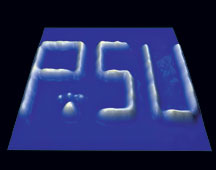
Scientists at Pennsylvania State University have demonstrated a technique for imaging and manipulating hydrogen atoms just below the surface of a palladium crystal (Proc. Natl. Acad. Sci. USA 2005, 102, 17907). Hydrogen interactions with precious metals are of key importance in hydrogen-storage applications, fuel cells, and other areas. In particular, subsurface H atoms have been fingered as intermediates in hydrogenation reactions on Pd, but until now the species has not been observed directly. By treating a Pd crystal with hydrogen at elevated temperature and pressure, E. Charles H. Sykes, Paul S. Weiss, and coworkers prepare samples in which a small amount of H atoms are absorbed into the crystal's bulk. Then, by applying voltage pulses to select positions on the crystal surface with a scanning tunneling microscope tip, the team induces the atoms to accumulate at stable sites just below the top Pd layer, where they can form patterns of lines measuring just a few nanometers wide, as shown.
GPCR assay monitors cell shape changes
G-protein-coupled receptors (GPCRs) are an important class of drug targets for a range of diseases. Most cellular screens for GPCRs require labeling or the use of reporter proteins. A team of scientists led by Yama A. Abassi of ACEA Biosciences has described a new noninvasive, label-free assay for GPCRs. This assay takes advantage of the fact that ligand binding to GPCRs induces a shape change in cells because the GPCRs are coupled to the actin cytoskeleton (Anal. Chem. 2006, 78, 35). Electronic cell sensor arrays measure changes in cell-electrode impedance caused by the cells changing shape when the GPCRs bind their ligands. The researchers demonstrate the assay with cells expressing recombinant receptors as well as cells expressing endogenous levels of histamine, calcitonin, or ??-opioid receptors. Incubation of the cells with receptor agonists led to dose-dependent changes in the impedance. The team can use the assay to screen both agonists and antagonists of GPCRs involved in different signaling pathways.
Taking a systems biology approach to apoptosis
A systems biology approach has uncovered previously unknown molecular mechanisms that link intracellular signaling to apoptosis, a type of cell death. A team of MIT researchers led by biologist Michael B. Yaffe developed a mathematical model to connect cellular signals and responses in a way that responses could be predicted from molecular signaling patterns (Science 2005, 310, 1646). Their apoptosis model is built from interactions of tumor necrosis factor (TNF, apoptosis protein) with epidermal growth factor or insulin, both of which inhibit apoptosis, but the model also reveals the contributions of other proteins. The original 660-dimension model could be simplified to a pair of orthogonal axes—a "stress-apoptosis axis" and a "survival axis"—that together provide a "molecular basis set for the signaling network that controls apoptosis." Using the model, Yaffe and coworkers discovered how circuits activated by the signaling proteins interleukin-1α and transforming growth factor-α cooperate with TNF in apoptosis. IL-1α serves as a positive-feedback loop, whereas TGF-α can send both pro- and antiapoptotic signals, depending on the circumstances.
Sea urchin's unforeseen molecular diversity
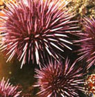
When biologist L. Courtney Smith of George Washington University and her colleagues exposed purple sea urchins in the laboratory to a bacterial fragment, the immune systems of the little spiny balls of marine life got busy. So busy, she finds, that she suspects she's uncovered a previously unknown arena of molecular diversity. Using DNA arrays to determine which genes turn on in macrophage-like cells in the exposed sea urchins, the researchers discovered what they estimate to be about 100 closely related proteins (Physiol. Genomics 2005, 22, 33). What's tantalizing about them, Smith says, is that their sequences are subtly variable in a way reminiscent of antibodies, the famously diverse megafamily of proteins in vertebrate immune systems. What's more, the newly found proteins appear to be encoded by mix-and-match genetic segments, a versatile arrangement that could foster a huge amount of sequence diversity. The discovery could help explain how invertebrates, whose evolutionary lineage predates the emergence of antibodies, have been able to ward off pathogenic challenges for so long, notes Sam Loker of the University of New Mexico, Albuquerque.
Secrets of toucan beak revealed
Using electron microscopy, the researchers found that the exterior of the beak is made up of overlapping tiles of keratin, the sulfur-containing fibrous protein that makes up hair, fingernails, and horn. The interior of the beak is constructed of a rigid foam (shown) made of a network of calcium-rich bony fibers connected by membranes. The membranes are similar in composition to keratin.
Meyers was surprised by the beak's ability to absorb high-energy impacts. Its structure could serve as the inspiration for automotive panels that could protect passengers in crashes and could also be used for ultralight aircraft components, according to the researchers.
Advertisement
You might also like...


The power is now in your (nitrile gloved) hands
Sign up for a free account to get more articles. Or choose the ACS option that’s right for you.
Already have an ACS ID? Log in
Join the conversation
Contact the reporter
Submit a Letter to the Editor for publication
Engage with us on Twitter