Advertisement
Grab your lab coat. Let's get started
Welcome!
Welcome!
Create an account below to get 6 C&EN articles per month, receive newsletters and more - all free.
It seems this is your first time logging in online. Please enter the following information to continue.
As an ACS member you automatically get access to this site. All we need is few more details to create your reading experience.
Not you? Sign in with a different account.
Not you? Sign in with a different account.
ERROR 1
ERROR 1
ERROR 2
ERROR 2
ERROR 2
ERROR 2
ERROR 2
Password and Confirm password must match.
If you have an ACS member number, please enter it here so we can link this account to your membership. (optional)
ERROR 2
ACS values your privacy. By submitting your information, you are gaining access to C&EN and subscribing to our weekly newsletter. We use the information you provide to make your reading experience better, and we will never sell your data to third party members.
Analytical Chemistry
Inside View
Technique provides high-resolution images of object interiors
by Carrie Arnold
July 21, 2008
| A version of this story appeared in
Volume 86, Issue 29

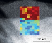
A NEW HIGH-RESOLUTION imaging technique bridges the gap between two different types of microscopy. The technique allows scientists to view a wide variety of electronic and biological samples at higher resolution and to visualize specimen interiors, which wasn't possible with the earlier methods.
The new technique, scanning X-ray diffraction microscopy (SXDM), combines the high resolution of coherent diffractive imaging (CDI) and the ease of sample preparation of scanning transmission X-ray microscopy (STXM). According to its developers, SXDM makes possible "imaging of the finest structures in state-of-the-art electronics devices or the macromolecular assemblies in organic tissues" (Science 2008, 321, 379).
CDI creates images by extrapolating structural information from the diffraction patterns of X-ray beams as they strike the specimen. This technique can produce high-resolution images, but image reconstruction and specimen preparation can be very difficult, and the range of accessible samples is limited to specimens completely isolated on a clean surface.
STXM forms images by measuring the transmitted intensity of an X-ray beam as it scans a sample. Although STXM allows the use of a much wider variety of specimens, its image resolution is limited by the size of the X-ray beam's focal spot.
Now, postdoc Pierre Thibault, assistant professor Franz Pfeiffer, and coworkers in the physics department at the Paul Scherrer Institut, in Villigen, Switzerland, have developed SXDM, an imaging and data reconstruction technique, to address the specimen applicability and resolution restrictions of the earlier techniques.
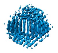
In their SXDM study, the researchers used a scanning beam of high-energy X-rays (as in STXM) and a high-speed detector to analyze a specimen containing a buried nanostructure. The team then examined the resulting X-ray diffraction patterns by the CDI method and reconstructed the image over a wide area of the sample. High-energy "X-rays can penetrate several tens of microns of material and can therefore image, in a noninvasive way, a buried nanostructure otherwise impossible to see," Thibault says.
These results, says physicist Chris Jacobsen of Stony Brook University, in New York, "show an impressive advance in spatial resolution and ease of reconstruction." Proposed uses for SXDM include investigating the interiors of superconductors and subcellular structures.


Join the conversation
Contact the reporter
Submit a Letter to the Editor for publication
Engage with us on Twitter