Advertisement
Grab your lab coat. Let's get started
Welcome!
Welcome!
Create an account below to get 6 C&EN articles per month, receive newsletters and more - all free.
It seems this is your first time logging in online. Please enter the following information to continue.
As an ACS member you automatically get access to this site. All we need is few more details to create your reading experience.
Not you? Sign in with a different account.
Not you? Sign in with a different account.
ERROR 1
ERROR 1
ERROR 2
ERROR 2
ERROR 2
ERROR 2
ERROR 2
Password and Confirm password must match.
If you have an ACS member number, please enter it here so we can link this account to your membership. (optional)
ERROR 2
ACS values your privacy. By submitting your information, you are gaining access to C&EN and subscribing to our weekly newsletter. We use the information you provide to make your reading experience better, and we will never sell your data to third party members.
Analytical Chemistry
Single Molecules Imaged In Living Mammalian Cell Nuclei
Fluorescence technique illuminates proteins in real time
by Jyllian Kemsley
March 25, 2013
| A version of this story appeared in
Volume 91, Issue 12
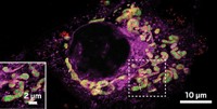
The ability to track individual biomolecules in live cells allows scientists to better understand the interactions between, for example, two proteins or a protein and DNA. Although methods exist to image single biomolecules in bacterial cells, imaging them in larger mammalian cells is challenging. Harvard University’s X. Sunney Xie and colleagues have now developed a fluorescence microscopy technique, called reflected light sheet microscopy, that illuminates proteins in the nucleus of living mammalian cells (Nat. Methods, DOI: 10.1038/nmeth.2411). The technique involves reflecting a thin sheet of light off a mirror—a disposable atomic force microscope cantilever coated with aluminum—so that the sheet slices horizontally through the cell and its nucleus. With the method, the team can detect fluorescently tagged proteins with nanometer spatial and millisecond time accuracy. In addition, the imaging can conveniently be carried out in the same commercially available glass-bottom culture dishes that are used to grow the cells. Xie and coworkers demonstrated the technique by imaging different transcription factors and their interactions with DNA and each other.


Join the conversation
Contact the reporter
Submit a Letter to the Editor for publication
Engage with us on Twitter